Cholesteatoma for NEET PG: Safe vs Unsafe CSOM, Complications and Surgery
Reflex · 3 Nov 2026 · 10 min read
Last updated: 3 Nov 2026
On this page
Cholesteatoma is the reason ENT textbooks split chronic suppurative otitis media (CSOM) into a safe type and an unsafe type. The two look alike when a patient first describes a discharging ear. The difference is that cholesteatoma erodes bone, and bone erosion is what opens the road to the facial nerve, the labyrinth and the brain.
This guide follows one reasoning chain: how to tell the types apart, why cholesteatoma forms, what it damages, how it is imaged, and how surgeons choose between canal wall up and canal wall down operations.
Key Takeaways
Safe (tubotympanic) CSOM has a central perforation and few serious complications. Unsafe (atticoantral) CSOM has an attic or posterosuperior marginal perforation and carries serious risk, usually because of cholesteatoma.
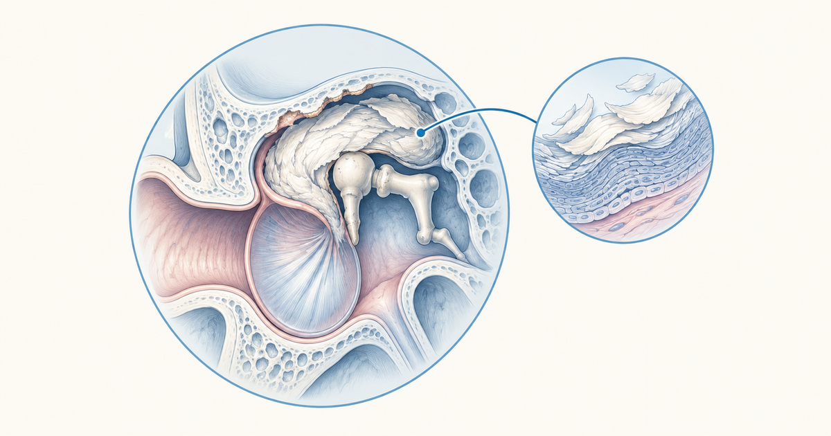
Cholesteatoma is keratinising stratified squamous epithelium (the matrix) sitting in the middle ear or mastoid, with inflammatory granulation tissue (the perimatrix) around it.
Most accepted theory: invagination of a retraction pocket. No single theory explains every case.
Complications split into intracranial (meningitis, brain abscess, sigmoid or lateral sinus thrombosis, petrositis) and extracranial or intratemporal (mastoiditis, facial palsy, labyrinthitis and fistula, postauricular and Bezold abscess).
HRCT shows bone erosion. MRI with diffusion-weighted imaging (DWI) shows the cholesteatoma itself.
Canal wall up keeps the canal wall but has more residual and recurrent disease. Canal wall down lowers recurrence but leaves an open cavity needing lifelong care.
Safe vs Unsafe CSOM: What Separates the Two Types?
The deciding feature is the site of the perforation and whether cholesteatoma is present. StatPearls describes the tubotympanic type as "benign", with a central, anteroinferior perforation and few serious complications. The atticoantral type is called "malignant" or the "danger type", with attic and posterosuperior perforations and a serious risk of complications. The commonly used labels are safe and unsafe CSOM.
The word "malignant" here means dangerous, not cancerous. Cholesteatoma is not a cancer, but it destroys bone as it expands.
| Feature | Safe (tubotympanic) CSOM | Unsafe (atticoantral) CSOM |
|---|---|---|
| Perforation | Central, anteroinferior | Marginal or attic, posterosuperior |
| Discharge | Mucopurulent | Persistent or recurrent, often foul, may be painless |
| Granulation or polyp | Possible | Polyp suggests cholesteatoma |
| Complication risk | Few serious complications | Serious risk |
| Hearing loss | Conductive | Conductive, may become sensorineural if the labyrinth is involved |
Sources: StatPearls CSOM, StatPearls cholesteatoma.
In one single-centre series of 1,146 cases, perforations were marginal or attic in 73.6% and central in 13.3% (Epidemiology of 1,146 cases, Braz J Otorhinolaryngol). Treat this as supporting detail from one centre, not a population figure.
Clinical Features of Each Type
In CSOM, patients have mucopurulent, sometimes foul otorrhoea and a central or marginal perforation. Granulation tissue or an aural polyp may be present. The hearing loss is conductive, so Rinne is negative and Weber lateralises to the affected ear. If you want to practise reading these results, see our guide to the Rinne and Weber tests.
In cholesteatoma, the discharge is painless, persistent or recurrent and foul. Otoscopy may show a white pearly mass in a posterosuperior retraction pocket. Vertigo suggests a labyrinthine fistula. Facial palsy occurs in about 1% to 3.4% of cases (StatPearls).
A granular polyp in a discharging ear should be treated as cholesteatoma until proven otherwise. This advice comes from a single review, so use it as a working rule rather than a hard criterion (Cureus 2022 review).
What Is Cholesteatoma and How Common Is It?
Cholesteatoma is a collection of keratinising stratified squamous epithelium in the middle ear or mastoid. The epithelium forms the matrix. The surrounding inflammatory granulation tissue is the perimatrix (Braz J Otorhinolaryngol 2005).
The middle-ear form affects about 6 to 9 per 100,000 people in developed countries. The male to female ratio is about 1.4:1, and about 85% of cases are unilateral (StatPearls; ESHNR imaging paper, Eur Radiol 2025).
Congenital vs Acquired
Congenital cholesteatoma makes up 4% to 24% of cases and arises from epithelial rests behind an intact drum. The Levenson criteria require an intact pars tensa and pars flaccida, no history of perforation and no prior ear surgery (Cureus 2022 review).
Acquired cholesteatoma is divided into two groups:
- Primary: arises from a retraction pocket.
- Secondary: arises in relation to a perforation.
About 80% of acquired cases start from the pars flaccida and about 20% from the pars tensa (ESHNR). The Tos classification names three sites: attic, tympanic sinus and pars tensa (Braz J Otorhinolaryngol 2005).
How Does Cholesteatoma Form? Pathogenesis Theories
The retraction pocket (invagination) theory is the most widely accepted, but no single theory explains every case. A 2016 review in Clinical and Experimental Otorhinolaryngology lists these theories:
| Theory | Proposed mechanism | Attributed to |
|---|---|---|
| Invagination (retraction) | Drum retracts into a pocket that traps keratin | Wittmack, 1933 (most accepted) |
| Basal cell hyperplasia | Epithelial cones grow through the basement membrane | Lange 1925, Ruedi 1978 |
| Immigration | Squamous epithelium migrates in through a perforation | Habermann and Bezold, 1890 |
| Metaplasia | Middle-ear mucosa turns into squamous epithelium | von Troltsch and Wendt (weakly supported) |
| Implantation | Epithelium is carried into the middle ear by injury or surgery | Iatrogenic or traumatic |
The Paparella continuum view fits the evidence better than any single theory. It treats cholesteatoma as multifactorial, so several mechanisms can operate in the same ear (Braz J Otorhinolaryngol 2005).
Why Does It Erode Bone?
Erosion comes from several sources working together: pressure from the expanding mass, enzymes such as collagenase, and cytokines including IL-1, TNF-alpha, PGE2 and matrix metalloproteinases. Bacterial biofilm and lipopolysaccharide add to the inflammation, and the RANKL/OPG pathway drives osteoclast activity (Clin Exp Otorhinolaryngol 2016).
This is why the ossicles suffer. Ossicular erosion is found in about 80% of cholesteatoma ears versus about 20% of non-cholesteatomatous COM.
Site matters too. Attic cholesteatoma leaves the chain intact in only 26% of cases, and pars tensa cholesteatoma erodes ossicles in about 90%. The long process of the incus is the most affected part (Braz J Otorhinolaryngol 2005; Epidemiology of 1,146 cases).
Complications of Cholesteatoma
Complications are what make unsafe CSOM unsafe. A useful way to learn them is to ask whether the disease has gone inward to the skull or outward through the temporal bone.
Memory aid (our own grouping, not a published mnemonic): inward = inside the skull (meninges, brain, venous sinuses, petrous apex); outward = through the temporal bone (mastoid, facial nerve, labyrinth, skin behind the ear).
| Group | Complications |
|---|---|
| Intracranial | Meningitis, brain abscess, sigmoid or lateral sinus thrombosis, petrositis (Gradenigo syndrome) |
| Extracranial or intratemporal | Mastoiditis, facial palsy, labyrinthitis, postauricular abscess, Bezold abscess |
Source: StatPearls CSOM.
Gradenigo syndrome is the triad of otitis, facial pain and ipsilateral abducens palsy. Fisch groups the same problems as intracranial (meningitis, abscess, venous sinus thrombosis) and temporal bone (mastoiditis, labyrinthine fistula, facial palsy, labyrinthitis, ossicular destruction). Saleh and Mills list lateral semicircular canal fistula, facial palsy, total sensorineural hearing loss, sinus thrombosis and intracranial invasion (Braz J Otorhinolaryngol 2005).
Clues That Point to a Specific Complication
Use the symptom to locate the structure:
- Vertigo in a cholesteatoma ear: think labyrinthine fistula.
- Facial weakness: think facial nerve erosion. It is the most common extracranial complication in one tertiary series (60%). For practice, see the facial nerve disorders questions.
- Swelling behind the ear: think subperiosteal abscess.
- Otitis with facial pain and sixth nerve palsy: think Gradenigo syndrome.
Labyrinthitis is usually linked to cholesteatoma or long-standing untreated otitis media, with bacteria reaching the inner ear more often through the round window than the oval window (StatPearls labyrinthitis).
How Often Do They Occur?
Complications are uncommon, but the stakes are high. A 20-year tertiary series found 55 complicated cases among 4,364 COM patients (1.26%), and only 0.21% had an intracranial complication.
Mortality in that series was 3.6% overall and 11.1% for intracranial complications. Subperiosteal abscess was more frequent in cholesteatoma ears (22.7% versus 6.1%). This is a single-centre series, so read it as an illustration of scale (Clinical Features and Outcomes of COM Complications, 20-year study, 2025).
Intracranial extension can reach the meninges, the brain or the venous sinuses.
Imaging of Cholesteatoma: HRCT and MRI
HRCT and MRI answer different questions. HRCT shows bone erosion and complications, including erosion of the scutum, lateral semicircular canal, tegmen and cochlea. MRI shows the lesion itself (ESHNR).
| Modality | What it shows |
|---|---|
| HRCT temporal bone | Bone erosion (scutum, lateral semicircular canal, tegmen, cochlea) and complications |
| MRI T2 | Cholesteatoma bright |
| MRI T1 | Low to intermediate signal, non-enhancing |
| DWI | Bright. Non-EPI or multishot sequences are optimal, since single-shot EPI is limited by artefacts |
| Gadolinium MRI | Used when complications are suspected |
Surgery: Canal Wall Up vs Canal Wall Down
Surgery removes the disease and aims for a safe, dry ear. The choice is between keeping the posterior canal wall (canal wall up) and removing it (canal wall down).
| Feature | Canal wall up | Canal wall down |
|---|---|---|
| Canal wall | Preserved | Removed, giving an open cavity |
| Cavity and aftercare | No open cavity | Open cavity needing lifelong care, water precautions, hearing-aid fitting problems |
| Recurrence | Higher risk | Lower |
| Residual disease in adults | 9% to 70% | 5% to 17% |
In children, residual disease is 16% to 54%, and the surgical approach has no significant influence. Sources: ESHNR; StatPearls.
Mastoid obliteration and canal reconstruction reduce the drawbacks of canal wall down. Atticotomy or atticoantrostomy suits small disease, endoscopic approaches are increasingly used, and subtotal petrosectomy is reserved for extensive disease.
When Do Surgeons Choose Canal Wall Down?
Canal wall down is often chosen during surgery when disease involves the sinus tympani or medial canal wall, or when there is a labyrinthine fistula. A patient with facial palsy before surgery usually gets canal wall down with facial nerve decompression (StatPearls). In the 20-year series, canal wall down was used in 61.8% of complicated cases and 77.8% of intracranial ones (20-year study of COM complications).
The canal wall down procedures include atticotomy, modified radical mastoidectomy and radical mastoidectomy (Cureus 2022 review).
For lateral sinus thrombosis, one expert letter describes canal wall down mastoidectomy with sinus puncture and the clot left in place, or clot removal with jugular ligation if there is perisinus abscess and sepsis. Treat this as opinion-level guidance (Eur Arch Otorhinolaryngol 2013).
Surgery for CSOM in General
Topical fluoroquinolone drops and aural toilet are first line. Surgery is indicated for otorrhoea persisting 4 to 6 weeks despite medical care, a non-healing perforation, cholesteatoma or coalescent mastoiditis. Typical organisms are Pseudomonas, Staphylococcus aureus, Proteus and Klebsiella (StatPearls CSOM).
Residual vs Recurrent Disease and Follow-Up
Residual disease means matrix was left behind at surgery. Recurrent disease means the retraction pocket has formed again (ESHNR).
Recurrence at 5 years is about 38% in children and 14% in adults (StatPearls). Follow-up with non-EPI DWI MRI is advised for at least 5 years after surgery. A second-look operation at 9 to 12 months is an older practice, used for canal wall up surgery.
Study Tips for Revising Cholesteatoma
Build the topic around three questions: where is the perforation, what has been eroded, and where has the disease spread? Draw the ear from memory, mark the attic and posterosuperior quadrant, then place each complication next to the structure that causes it. Rehearse the safe versus unsafe table until you can write it without looking.
To test yourself on this topic, work through the external and middle ear practice questions.
Conclusion
Cholesteatoma turns an ordinary discharging ear into a dangerous one because it destroys bone. Safe CSOM has a central perforation and mucosal disease, while unsafe CSOM has an attic or posterosuperior marginal perforation and a risk of intracranial and temporal bone complications. HRCT maps the erosion, DWI MRI finds the lesion, and surgery chooses between preserving the canal wall and accepting an open cavity for lower recurrence. Long follow-up matters because disease can return years later.
Cholesteatoma is also covered in the NEET PG question bank, where you can practise the same reasoning on stems.
Practise this on Reflex
Turn what you just read into recall with 14 years of tagged PYQs.
FAQ
Frequently asked questions
The questions aspirants ask most about this topic.
Safe (tubotympanic) CSOM has a central, anteroinferior perforation and few serious complications. Unsafe (atticoantral) CSOM has an attic or posterosuperior marginal perforation and carries a serious risk of complications, usually because cholesteatoma is present. StatPearls calls these the benign and danger types.
The invagination or retraction pocket theory (Wittmack, 1933) is the most accepted. No single theory explains every case, so cholesteatoma is viewed as multifactorial.
Intracranial complications include meningitis, brain abscess, sigmoid or lateral sinus thrombosis and petrositis (Gradenigo syndrome). Extracranial or intratemporal complications include mastoiditis, facial palsy, labyrinthitis, postauricular abscess and Bezold abscess.
HRCT temporal bone shows bone erosion and complications. MRI shows the cholesteatoma itself: bright on T2 and DWI, low to intermediate on T1 and non-enhancing. Non-EPI or multishot DWI is optimal because single-shot EPI is limited by artefacts.
Canal wall up preserves the canal wall and gives better hearing and cosmetics but a higher risk of residual and recurrent disease. Canal wall down removes the wall, lowers recurrence, and leaves an open cavity that needs lifelong care, water precautions and may cause hearing-aid problems.
Residual disease results from incomplete removal of the matrix at surgery. Recurrent disease is the reformation of the retraction pocket after surgery.
Stay ahead in your preparation
Get expert tips, exam updates, and high-yield insights delivered straight to your inbox.
No spam. Unsubscribe anytime.
More in Subject Notes
View all → ENT
Epistaxis for NEET PG: Little's Area, Woodruff's Plexus, and the Management Ladder
 ENT
Rinne and Weber Test for NEET PG: Reading the Hearing Loss Pattern Correctly
The two tests are simple individually; the exam rewards reading them together and landing on conductive versus sensorineural without hesitation. The combined pattern, why the direction is easy to get backwards, and how audiometry confirms it.
 Physiology
Cardiac Cycle for NEET PG: A Phase-by-Phase Walkthrough of the Wiggers Diagram
A phase-by-phase walkthrough of the cardiac cycle on the Wiggers diagram, with valve states, heart sounds, ECG timing and JVP waves in revision tables.
 Biochemistry
Vitamin Deficiency Diseases for NEET PG: A Vitamin-by-Vitamin Table of Classic Signs
A revision guide to vitamin deficiency diseases: fat-soluble and water-soluble vitamins in tables, with classic signs, lab clues, treatment, and the look-alike pairs that cause confusion.