Rinne and Weber Test for NEET PG: Reading the Hearing Loss Pattern Correctly
By Dr. Utsav Bhattacherjee, MBBS, MBA · 25 Aug 2026 · 9 min read
Last updated: 23 Sept 2026
On this page
ENT has a short syllabus relative to Medicine or Surgery, and Rinne and Weber testing is close to the center of it — a bedside tool that shows up constantly in both theory questions and clinical vignettes. The two tests are simple individually; the exam rewards being able to read them together and land on conductive versus sensorineural hearing loss without hesitation.
How Each Test Works
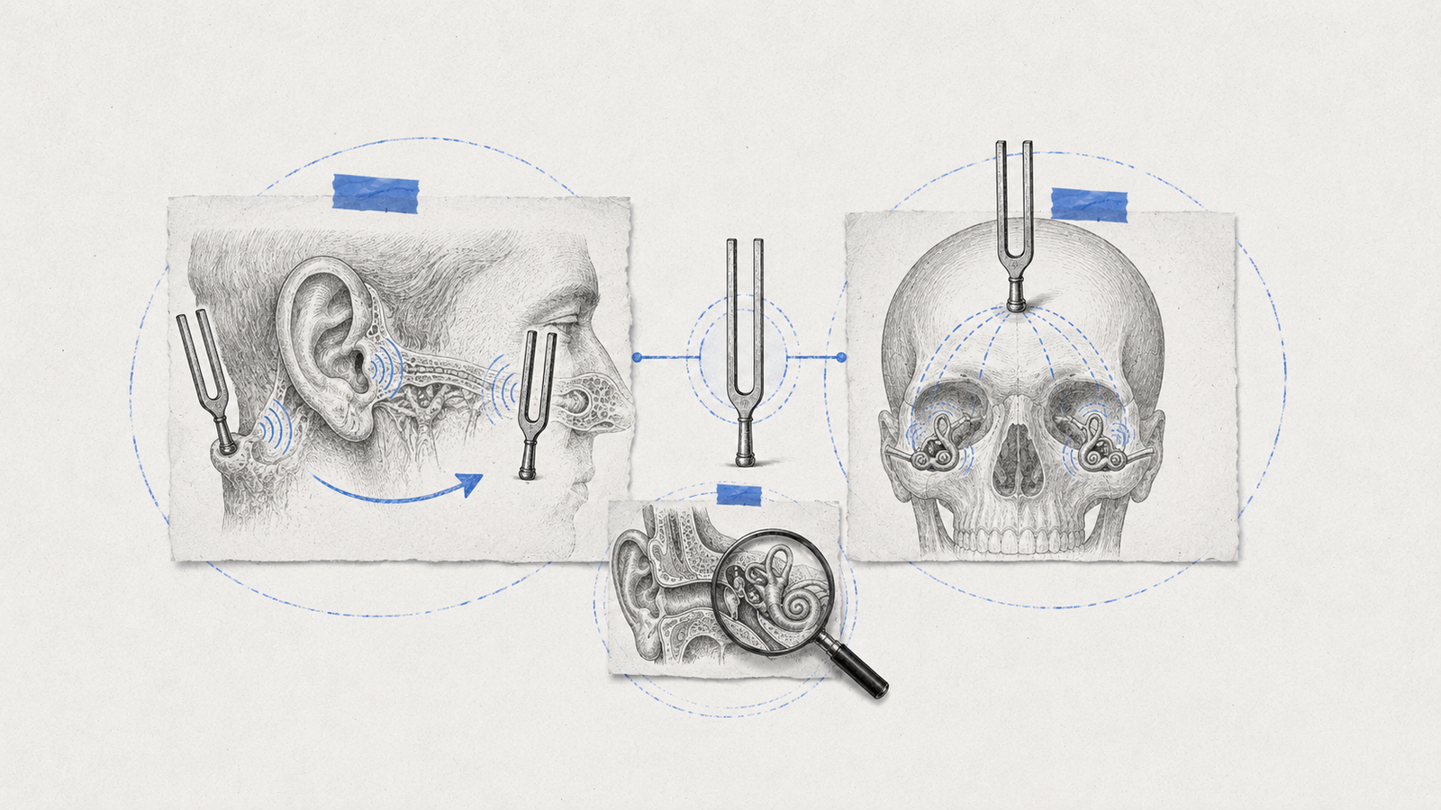
Rinne test compares air conduction (AC) to bone conduction (BC) within the same ear. A vibrating tuning fork is placed on the mastoid process until the sound is no longer heard, then moved next to the ear canal. Normally, air conduction outlasts bone conduction — a Rinne positive result (AC greater than BC). If bone conduction is heard longer or louder than air conduction, that's Rinne negative (BC greater than AC).
Weber test places a vibrating tuning fork at the midline of the skull — typically the forehead or vertex — and the patient reports which side, if any, the sound is louder in. This is called lateralization, and unlike Rinne, it compares the two ears against each other rather than testing one ear's own two conduction pathways.
Reading the Combined Pattern
| Condition | Rinne (affected ear) | Weber lateralization |
|---|---|---|
| Normal hearing | Positive (AC greater than BC) | Midline, no lateralization |
| Conductive hearing loss | Negative (BC greater than AC) | Lateralizes TO the affected ear |
| Sensorineural hearing loss | Positive (AC greater than BC, though both reduced) | Lateralizes AWAY FROM the affected ear, toward the better ear |
Why the Direction Is Easy to Get Backwards
The counterintuitive part is conductive loss: Weber lateralizes toward the bad ear, not away from it. A conductive problem — fluid, wax, or a fixed ossicular chain — blocks ambient background noise from reaching that ear, which makes bone-conducted sound seem relatively louder there by comparison, since there's less competing external noise masking it. In sensorineural loss, the cochlea or auditory nerve itself is damaged, so that ear is simply worse at perceiving sound altogether regardless of pathway — the healthy ear picks up the vibration better, and lateralization goes toward the normal side instead.
A Worked Example
A patient has fluid behind the left eardrum from otitis media with effusion. Expect: Rinne negative in the left ear (bone conduction outlasts air conduction, since the fluid blocks the air-conduction pathway specifically), and Weber lateralizing to the left ear — the classic conductive pattern, where the affected ear paradoxically "wins" the Weber test.
Now compare a patient with left-sided sudden sensorineural hearing loss: Rinne stays positive in the left ear (air conduction still outlasts bone conduction, just both are reduced overall), and Weber lateralizes to the right — the unaffected side — since the left cochlea is less able to perceive the vibration regardless of how it arrives.
Causes Behind Each Pattern
Conductive hearing loss arises from a problem anywhere along the pathway before sound reaches the cochlea — cerumen (earwax) impaction, otitis media with effusion, otosclerosis (fixation of the stapes), tympanic membrane perforation, or ossicular chain discontinuity.
Sensorineural hearing loss arises from a problem in the cochlea or the auditory nerve pathway itself — presbycusis (age-related hearing loss), noise-induced hearing loss, ototoxic drugs (aminoglycosides, loop diuretics, and certain chemotherapy agents are the classic culprits), Meniere's disease, and vestibular schwannoma (acoustic neuroma) affecting the nerve directly.
Otosclerosis deserves a specific mention among the conductive causes, since it's a favorite in exam vignettes: it fixes the stapes footplate to the oval window, progressively blocking sound transmission, and classically presents in a young adult with progressive hearing loss and a family history — sometimes accompanied by tinnitus. It's also a case where surgical management (stapedectomy) can be genuinely curative, unlike most sensorineural causes, which is part of why the conductive-versus-sensorineural distinction has real clinical stakes beyond the exam itself.
Confirming the Pattern With Audiometry
Tuning fork tests are a fast bedside screen, but pure tone audiometry is what confirms and quantifies the finding. On an audiogram, conductive hearing loss shows a characteristic air-bone gap — bone conduction thresholds stay relatively normal while air conduction thresholds are elevated, visually separating the two curves. Sensorineural hearing loss shows both air and bone conduction thresholds reduced roughly together, with no meaningful gap between them, since the deficit lies beyond where the air-versus-bone distinction matters. Recognizing this pattern on an audiogram is really the same underlying logic as the tuning fork tests, just measured with more precision.
Grading Hearing Loss by Severity
Once a hearing loss is identified and classified as conductive or sensorineural, audiometry also grades it by severity, based on the hearing threshold in decibels (dB):
| Grade | Threshold |
|---|---|
| Normal | Up to 25 dB |
| Mild | 26–40 dB |
| Moderate | 41–55 dB |
| Moderately severe | 56–70 dB |
| Severe | 71–90 dB |
| Profound | Above 90 dB |
This grading matters clinically because it drives management — a mild conductive loss from wax impaction is managed very differently from a profound sensorineural loss being evaluated for a cochlear implant, even though both might show up in a question stem simply as "hearing loss" before the grading detail is introduced.
Absolute Bone Conduction and Schwabach Tests
A third tuning fork test worth knowing alongside Rinne and Weber: the Absolute Bone Conduction (ABC) test compares the patient's bone conduction, with the external ear canal occluded, against the examiner's. It's normal in conductive hearing loss, since the cochlea and nerve pathway are intact — the problem is purely mechanical, upstream of where bone conduction bypasses it. It's reduced in sensorineural hearing loss, since the deficit lies in the cochlea or nerve itself, which bone conduction can't route around. Combined with Rinne and Weber, ABC is occasionally used as a tie-breaker in an ambiguous presentation, though in practice Rinne and Weber together are usually sufficient to reach a confident answer.
The Schwabach test compares the patient's own bone conduction duration to the examiner's (assumed normal) bone conduction, using the same tuning fork placed on the mastoid of each in turn. A prolonged Schwabach (the patient hears the bone-conducted sound longer than the examiner does) suggests conductive hearing loss, since bone conduction is relatively enhanced when the competing air-conduction pathway is blocked. A shortened Schwabach suggests sensorineural hearing loss, since the patient's cochlea itself perceives bone-conducted sound for less time than a normal ear would. It's tested less frequently than Rinne and Weber, but it's a useful cross-check when a question wants to confirm you understand the underlying logic rather than just the two most familiar tests.
Pattern-reading questions like this are exactly what fast, timed practice sharpens. For a related lab-and-clinical-pattern topic covered the same way, our guide to jaundice types works through the same kind of paired-findings recognition, and for how ENT fits into your overall exam weightage, see our subject-wise weightage breakdown.
Practise this on Reflex
Turn what you just read into recall with 14 years of tagged PYQs.
FAQ
Frequently asked questions
The questions aspirants ask most about this topic.
Bone conduction is heard longer or louder than air conduction in that ear — the hallmark of conductive hearing loss.
Away from the affected ear, toward the better-hearing ear.
Toward the affected ear — the side with the conductive problem, counterintuitively.
Conductive loss shows Rinne negative and Weber lateralizing toward the bad ear. Sensorineural loss shows Rinne positive (though reduced) and Weber lateralizing toward the good ear.
Conductive hearing loss — bone conduction thresholds stay near-normal while air conduction thresholds are elevated, creating a visible gap between the two curves.
Conductive: cerumen impaction, otitis media with effusion, otosclerosis, tympanic membrane perforation. Sensorineural: presbycusis, noise exposure, ototoxic drugs, Meniere's disease, and vestibular schwannoma.
About the author
Stay ahead in your preparation
Get expert tips, exam updates, and high-yield insights delivered straight to your inbox.
No spam. Unsubscribe anytime.
More in Subject Notes
View all → ENT
ENT