FAST Scan for NEET PG: The Four Windows, eFAST, and What a Positive Result Means
By Dr. Utsav Bhattacherjee, MBBS, MBA · 5 Oct 2026 · 10 min read
Last updated: 8 Oct 2026
On this page
FAST questions rarely test technique. They test what a positive or negative scan should make you do next. Learn four windows, one extension and one decision tree, and most questions can be answered without ever seeing the images.
What FAST Is and Why It Exists
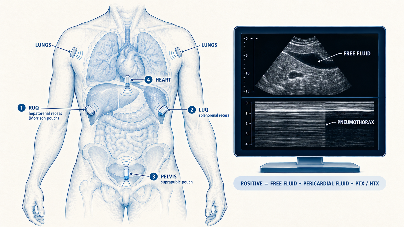
FAST stands for Focused Assessment with Sonography for Trauma. It is a rapid bedside ultrasound that looks for free fluid in the pericardial sac and the peritoneal cavity, and in a trauma patient that fluid is presumed to be blood. It answers one question, whether there is free fluid, and it does not tell you which organ is injured. Its strengths are practical: it takes only a few minutes, it can be done during resuscitation without moving the patient, it can be repeated as often as needed, it uses no radiation and it is safe in pregnancy. A low-frequency probe, roughly 2 to 5 MHz, gives the depth needed for the abdomen.
Where FAST Fits in the Primary Survey
In the ATLS sequence, FAST is an adjunct to the primary survey, used while assessing circulation in a patient who is hypotensive or has an unreliable examination, for example because of a head injury or intoxication. The usual indications are blunt abdominal trauma with hypotension, penetrating torso trauma and suspected cardiac injury. There is no absolute contraindication, but FAST must never delay resuscitation or transfer to theatre when the need for laparotomy is already obvious.
The Four Standard Windows
| Window | Probe position | What you look for | Why it matters |
|---|---|---|---|
| Pericardial | Below the xiphoid, aimed towards the left shoulder | A black stripe around the heart | Haemopericardium and tamponade |
| Right upper quadrant | Right mid-axillary line, lower ribs | Fluid in the hepatorenal space (Morison's pouch) and around the liver tip | The most commonly positive window |
| Left upper quadrant | Left posterior axillary line, lower ribs | Fluid between the spleen and kidney and above the spleen | Splenic injury |
| Suprapubic (pelvic) | Above the pubic symphysis, using the bladder as a window | Fluid behind the bladder or in the pouch of Douglas | The pelvis collects fluid from elsewhere in the abdomen |
Free fluid is anechoic, so it appears black, and the core skill is spotting a black stripe where there should be none. Fresh blood is anechoic, but clotted blood can look echogenic, which is one way a real bleed is missed. Small amounts of pelvic fluid in women can be physiological, so the result is always read in its clinical context.
eFAST: Adding the Chest
The extended FAST adds views of both sides of the anterior chest to look for pneumothorax and haemothorax. In a normal lung the pleural line slides with breathing, called lung sliding, and on M-mode this gives the granular seashore sign. In pneumothorax, lung sliding is absent and M-mode shows the barcode (stratosphere) sign. The lung point, where sliding reappears at the edge of the collapsed lung, is highly specific. Haemothorax appears as a black collection above the diaphragm, and the vertebral column becomes visible above the diaphragm (the spine sign).
Pericardial Fluid or Pleural Fluid?
A common trap on the cardiac view is telling a pericardial effusion from a left pleural effusion. The descending thoracic aorta is the landmark: pericardial fluid lies in front of it, and pleural fluid lies behind it. In tamponade, right atrial and right ventricular collapse appear and the inferior vena cava looks plethoric (dilated and non-collapsing). In trauma, haemopericardium may contain clot and look echogenic, so a bright rather than black collection around the heart still counts.
Reading the Result: What Do You Do Next?
A FAST result matters only for how it changes the next step, and the logic follows the patient's haemodynamic state.
| Patient | FAST result | Next step |
|---|---|---|
| Blunt trauma, haemodynamically unstable | Positive | Emergency laparotomy |
| Blunt trauma, haemodynamically unstable | Negative | Look for other sources of shock (chest, pelvis, long bones, external bleeding) and repeat the scan |
| Blunt trauma, haemodynamically stable | Positive | Contrast CT of the abdomen to define the injuries |
| Blunt trauma, stable, with ongoing clinical concern | Negative | Serial examination, repeat FAST, CT as needed |
| Penetrating chest injury | Fluid around the heart | Urgent surgical exploration |
The most tested row is the second one. A negative FAST in an unstable patient does not rule out intra-abdominal bleeding, because small or early bleeds are missed, so the answer is to look for other sources of shock and to repeat the scan, not to reassure.
What FAST Cannot Do
FAST detects fluid, not injury. It can miss solid organ injuries that have not yet bled freely, retroperitoneal injuries (kidney, pancreas, duodenum), hollow viscus injuries and diaphragmatic injuries. It cannot say what the fluid is, so ascites, urine and old blood look much like fresh blood. It depends on the operator, and its views are degraded by obesity, subcutaneous emphysema and bowel gas. If possible, scan the pelvis before a urinary catheter is inserted, because a full bladder acts as an acoustic window and an empty one makes the view harder.
FAST, Peritoneal Lavage and CT Compared
| Test | Strength | Weakness |
|---|---|---|
| FAST | Immediate, bedside, repeatable and radiation-free; works during resuscitation | Operator-dependent; detects fluid, not organ injury; misses retroperitoneal and hollow viscus injury |
| Diagnostic peritoneal lavage | Sensitive for intraperitoneal blood and for hollow viscus injury | Invasive and not organ-specific; can lead to an unnecessary laparotomy; largely replaced by FAST |
| Contrast CT of the abdomen | Best for grading organ injury and showing the retroperitoneum | Needs a stable patient, transport and contrast; uses radiation |
Pitfalls on the Screen
- A fluid-filled stomach or bowel can mimic free fluid in the left upper quadrant, and the gallbladder can mimic it on the right.
- The epicardial fat pad can be mistaken for a small pericardial effusion.
- A decompressed bladder removes the window for the pelvic view.
- A small amount of physiological pelvic fluid in a woman can be mistaken for injury.
Exam Pearls
- FAST is a screening test for free fluid, not a test for organ injury.
- The hepatorenal space (Morison's pouch) is the most commonly positive window.
- Unstable blunt trauma with a positive FAST goes to the operating theatre, not to CT.
- A negative FAST in an unstable patient means look elsewhere and repeat, not reassure.
- Absent lung sliding with a lung point is pneumothorax on eFAST.
- Pericardial fluid lies in front of the descending aorta; pleural fluid lies behind it.
Penetrating Trauma and the Pericardial Window
In penetrating chest trauma near the heart, the pericardial view is the most important of the four. Fluid around the heart in an unstable patient with a precordial wound means haemopericardium until proven otherwise, and the answer is urgent surgical exploration without waiting for further imaging. Even a small collection matters in this setting. The pericardium is a closed space, and a small volume of acutely accumulated blood can compromise cardiac filling long before a chronic effusion of the same size would.
FAST in Children and in Pregnancy
Because FAST uses no radiation, it is especially useful in children and in pregnant women. In children most solid organ injuries are managed without surgery, so in a haemodynamically stable child a positive FAST leads to selective CT rather than automatic laparotomy. In pregnancy, ultrasound does not expose the fetus to radiation, so it is a sensible first test, but the enlarged uterus can make the scan harder to read, and a negative result should be interpreted with care.
How Vignettes Are Built
Check the haemodynamic state first, then the FAST result, then the mechanism. A man pulled from a road traffic collision with a blood pressure of 80/50, a rising pulse and free fluid in Morison's pouch needs a laparotomy, and CT is the wrong answer however tempting it looks. The same man with a normal blood pressure and the same finding needs a contrast CT to define the injury. A stab wound to the left chest with fluid around the heart on the subxiphoid view needs urgent surgical exploration. If the question stresses a retroperitoneal organ, think of FAST's blind spots.
For the wider imaging picture, including chest X-ray signs, CT head patterns and MRI sequences, see our radiology high-yield guide, and for how much of the paper Radiology carries, our subject-wise weightage breakdown.
Practise this on Reflex
Turn what you just read into recall with 14 years of tagged PYQs.
FAQ
Frequently asked questions
The questions aspirants ask most about this topic.
Focused Assessment with Sonography for Trauma. It is a rapid bedside ultrasound that looks for free fluid in the pericardial sac and the peritoneal cavity, which in a trauma patient is presumed to be blood.
The pericardial window, the right upper quadrant (hepatorenal space), the left upper quadrant (splenorenal space) and the suprapubic (pelvic) window.
It adds views of the anterior chest on both sides to detect pneumothorax (absent lung sliding, barcode sign on M-mode, lung point) and haemothorax (black fluid above the diaphragm).
Emergency laparotomy, not CT. The scan has already shown that there is free fluid, and the patient is too unstable to go to the scanner.
Small or early bleeds are missed, and FAST cannot see retroperitoneal injuries. The answer is to look for other sources of shock and repeat the scan.
Retroperitoneal injuries such as kidney, pancreas and duodenum, hollow viscus injuries, diaphragmatic injuries, and solid organ injuries that have not yet bled freely.
Pericardial fluid lies in front of the descending thoracic aorta on the cardiac view, while pleural fluid lies behind it.
About the author
Stay ahead in your preparation
Get expert tips, exam updates, and high-yield insights delivered straight to your inbox.
No spam. Unsubscribe anytime.
More in Subject Notes
View all → Radiology
Radiology for NEET PG 2026: X-Ray Signs, CT Patterns, and Complete Imaging Guide
Radiology is 3 to 6 questions of pure pattern recognition. Sixty-plus named signs, the TB patterns, tension pneumothorax and the extradural versus subdural distinction — the ones that actually repeat.
 Pediatrics
Kawasaki Disease for NEET PG: The Five Criteria, the 10-Day IVIG Window and the Coronary Risk
What counts as Kawasaki disease, why the 10-day IVIG window matters, how aspirin is used, and how coronary artery involvement is graded and followed.
 Pediatrics
Tetralogy of Fallot for NEET PG: One Septal Shift, Four Defects, and the Tet Spell
One shifted septum explains the four defects of Tetralogy of Fallot, the boot-shaped heart, the fading murmur and the stepwise management of a tet spell.
 Microbiology
Malaria Life Cycle for NEET PG: Species, Smear Morphology, Complications and Treatment
How the malaria parasite moves between mosquito and human, what separates the species on a smear, and how diagnosis, complications and treatment follow from the life cycle.