Radiology for NEET PG 2026: X-Ray Signs, CT Patterns, and Complete Imaging Guide
By Dr. Utsav Bhattacherjee, MBBS, MBA · 3 Aug 2026 · 20 min read
Last updated: 23 Sept 2026
On this page
Radiology contributes approximately 2 to 3 percent of the NEET PG paper — around 3 to 6 questions out of 180. These questions are almost exclusively pattern recognition: a described finding, a named sign, or a clinical scenario where you must identify the correct imaging modality or characteristic appearance. Radiology PYQs are highly predictable — the same signs, the same TB patterns, the same CT findings appear cycle after cycle.
This guide covers every high-yield Radiology topic with the depth NEET PG demands. The signs tables alone capture more than 60 sign-disease pairs tested in PYQs. For where Radiology sits against the rest of the paper, see the subject wise weightage — Radiology; for how to slot it into your timetable, see the NEET PG 2026 preparation strategy.
Before starting revision, use the NEET PG 2026 score calculator to track how your subject-wise accuracy maps to your projected rank. Radiology is one of the most efficiently studied subjects — 5 to 7 focused days of PYQ-based preparation can reliably get you every Radiology question.
Radiology rewards the same habit as the other sign-heavy subjects: learn the eponym, learn the disease, move on. If you are batching them, Dermatology for NEET PG 2026 works identically, and Anaesthesia for NEET PG 2026 covers the third of the small, high-repetition subjects.
The 14 Most-Tested Radiology Topics in NEET PG PYQs
| Rank | Topic | Frequency | Format |
|---|---|---|---|
| 1 | Named X-ray and CT signs — disease associations | Very High | Direct factual |
| 2 | Tuberculosis — primary, post-primary, miliary | Very High | Pattern recognition |
| 3 | Pneumothorax vs tension pneumothorax | Very High | Clinical scenario |
| 4 | Pleural effusion — X-ray findings and types | High | Direct factual |
| 5 | CT head — extradural vs subdural haematoma | High | Comparison MCQ |
| 6 | GI radiology — barium studies and named findings | High | Direct factual |
| 7 | Pulmonary embolism — Westermark sign, Hampton's hump | High | Direct factual |
| 8 | Cardiac radiology — cardiomegaly, boot-shaped heart | Moderate | Direct factual |
| 9 | Paediatric radiology — steeple, double bubble, thumb sign | Moderate | Clinical scenario |
| 10 | Silhouette sign — principle and applications | Moderate | Concept plus application |
| 11 | Ultrasound — high-yield findings | Moderate | Direct factual |
| 12 | MRI — named sequences and disease patterns | Moderate | Direct factual |
| 13 | Radiation physics — dose, units, safety | Low-Moderate | Direct factual |
| 14 | Interventional radiology — procedures and indications | Low-Moderate | Direct factual |
Chest X-Ray: A Systematic Approach
NEET PG clinical scenario questions often describe a chest X-ray finding, so reading the description correctly matters as much as knowing the signs.
| Step | What to Check |
|---|---|
| A — Airway | Trachea midline or deviated; carina angle |
| B — Breathing | Lung fields for opacification, lucency, consolidation, cavities, nodules, pleural lines |
| C — Cardiac | Heart size (cardiothoracic ratio), borders, shape |
| D — Diaphragm | Levels, costophrenic angles, free air beneath |
| E — Everything else | Bones, soft tissues, tubes, lines, foreign bodies |
The cardiothoracic ratio is normally 0.5 or less on a PA film taken in full inspiration. Above 0.5 indicates cardiomegaly. An AP film magnifies the cardiac silhouette, so the ratio is unreliable there.
The Complete Radiology Signs Reference
Chest X-Ray Signs
| Sign | Appearance | Condition |
|---|---|---|
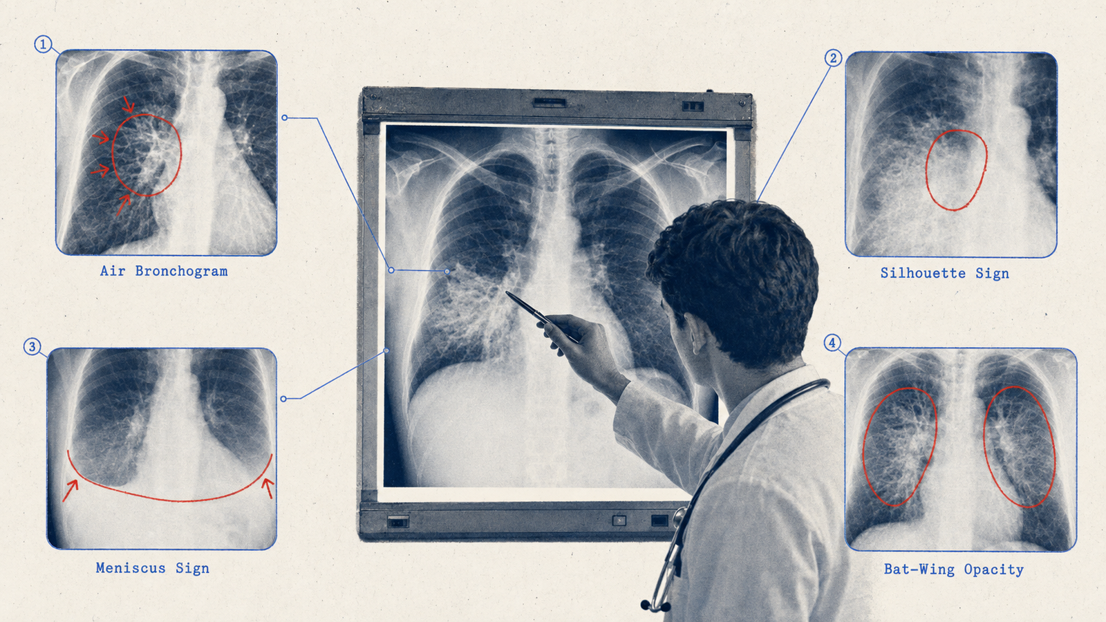
| Silhouette sign | Loss of the interface between two adjacent structures of the same density | Consolidation touching a cardiac border — right middle lobe loses the right heart border, lingula the left |
| Air bronchogram | Dark branching airways visible through white opacified lung | Consolidation — air in airways surrounded by fluid-filled alveoli |
| Meniscus sign | Concave upper border of fluid, highest peripherally | Pleural effusion |
| Vertical fluid line | Straight horizontal fluid level | Hydropneumothorax |
| Westermark sign | Focal oligaemia distal to an embolus | Pulmonary embolism |
| Hampton's hump | Peripheral wedge-shaped opacity based on the pleura | Pulmonary infarction from PE |
| Fleischner lines | Linear opacities representing atelectasis | Pulmonary embolism; post-operative atelectasis |
| Kerley B lines | Short horizontal peripheral lines perpendicular to the pleura | Pulmonary oedema; lymphangitis carcinomatosa |
| Bat wing pattern | Bilateral perihilar opacification sparing the periphery | Cardiogenic pulmonary oedema |
| Sail sign (thymic) | Triangular right mediastinal opacity | Normal thymus in infants |
| Sail sign (atelectasis) | Triangular opacity of a collapsed lobe | Right middle or left lower lobe collapse |
| Golden S sign | S-shaped curve from a collapsed upper lobe plus a central mass | Right upper lobe collapse from a hilar mass, usually bronchogenic carcinoma |
| Pleural cap | Opacity above the lung apex | Apical pleural thickening; Pancoast tumour; haemothorax |
| Cervicothoracic sign | A lesion disappearing above the clavicle is posterior; one still visible is anterior | Localising mediastinal masses |
| Tram-track lines | Parallel opacities from thickened bronchial walls | Bronchiectasis |
| Signet ring sign | Dilated airway larger than its adjacent pulmonary artery | Bronchiectasis |
| Tree-in-bud pattern | Centrilobular nodules with branching opacities | Endobronchial spread of tuberculosis |
| Halo sign | Ground-glass opacity surrounding a nodule | Invasive aspergillosis; early haemorrhagic nodule |
| Reverse halo (atoll) sign | Central lucency ringed by ground-glass or consolidation | Cryptogenic organising pneumonia; fungal infection |
| Galaxy sign | Large central opacity with satellite nodules | Pulmonary sarcoidosis |
| Eggshell calcification | Calcification confined to the rim of hilar nodes | Silicosis; sarcoidosis; treated lymphoma |
| Popcorn calcification | Irregular calcification throughout a nodule | Pulmonary hamartoma |
| Air crescent sign | Crescent of air around a mass within a cavity | Aspergilloma |
| Monod sign | The fungal ball moves with position change | Aspergilloma |
| Water lily sign | Undulating membranes within a cyst | Pulmonary hydatid — collapsed endocyst |
| Cannon ball metastases | Multiple large rounded lung opacities | Choriocarcinoma; renal cell carcinoma; testicular germ cell tumour |
| Boot-shaped heart | Upturned apex with reduced pulmonary vascularity | Tetralogy of Fallot |
| Egg on its side | Narrow superior mediastinum with a large oval heart | D-transposition of the great arteries |
| Box-shaped heart | Markedly enlarged globular heart | Ebstein anomaly; massive pericardial effusion |
| Snowman heart | Large supracardiac shadow | Supracardiac total anomalous pulmonary venous connection |
| Rib notching | Inferior rib erosion from collateral arteries | Coarctation of the aorta |
| Figure 3 sign | Aortic knob with pre- and post-stenotic dilatation | Coarctation of the aorta |
Pneumothorax and Tension Pneumothorax
A simple pneumothorax shows a visible pleural line with absent lung markings beyond it, partial collapse on the affected side, and a midline or minimally deviated trachea with no mediastinal shift.
Tension pneumothorax is different, and it is an emergency.
| X-Ray Feature | Direction | Significance |
|---|---|---|
| Tracheal deviation | Away from the affected side | Trapped air pushing the mediastinum |
| Mediastinal shift | Away from the affected side | Compressive effect |
| Diaphragm | Depressed and flattened on the affected side | Air expanding downward |
| Lung | Completely collapsed to a white opacity at the hilum | Total collapse |
| Contralateral lung | Compressed by the shifted mediastinum | Reduced ventilation |
| Heart | Shifted away from the pneumothorax | May mimic dextrocardia |
The clinical triad is absent breath sounds on the affected side, tracheal deviation away from it, and hypotension. Treatment is immediate needle decompression at the second intercostal space in the midclavicular line. Do not wait for X-ray confirmation.
Paediatric Signs
| Sign | Appearance | Condition |
|---|---|---|
| Steeple sign | Subglottic tracheal narrowing on AP neck film | Viral croup |
| Thumb sign | Swollen epiglottis on lateral neck film | Acute epiglottitis |
| Double bubble sign | Two air bubbles, stomach and duodenum | Duodenal atresia; annular pancreas |
| Triple bubble sign | Three air bubbles | Jejunal atresia |
| Ground glass abdomen | Diffuse fine granular opacity in a neonate | Meconium ileus; early necrotising enterocolitis |
| Soap bubble appearance | Multiple small bubbles in the right lower quadrant | Meconium ileus |
| Football sign | Large rounded abdominal lucency | Massive pneumoperitoneum |
| Bird of prey sign | Beak-like narrowing of the sigmoid | Sigmoid volvulus |
| Coffee bean sign | Large omega-shaped bowel loop | Sigmoid volvulus |
Gastrointestinal Signs
| Sign | Imaging | Appearance | Condition |
|---|---|---|---|
| String sign of Kantor | Barium meal | String-like narrowing of terminal ileum | Crohn's disease |
| Lead pipe colon | Barium enema | Loss of haustra, rigid smooth colon | Long-standing ulcerative colitis |
| Cobblestone pattern | Barium | Deep ulcers alternating with oedematous mucosa | Crohn's disease |
| Rose-thorn ulcers | Barium | Spike-like projections from the bowel wall | Crohn's disease |
| Collar-stud ulcer | Barium | Flask-shaped ulcer penetrating the mucosa | Ulcerative colitis |
| Apple core lesion | Barium or CT | Circumferential annular constricting lesion | Colonic carcinoma |
| Bird's beak | Barium swallow | Smooth tapering of the distal oesophagus | Achalasia cardia |
| Corkscrew oesophagus | Barium swallow | Segmental spasms | Diffuse oesophageal spasm |
| Rigler sign | Plain film | Both sides of the bowel wall visible | Pneumoperitoneum |
| Psoas shadow loss | Plain film | Loss of the psoas outline | Retroperitoneal abscess, haematoma or tumour |
| Whirlpool sign | CT | Swirling mesenteric vessels | Midgut or caecal volvulus |
| Accordion sign | CT | Thickened colonic haustra | Pseudomembranous colitis |
| Target sign | CT or ultrasound | Concentric layers around bowel wall | Intussusception |
| Mercedes-Benz sign | Plain film or CT | Y-shaped lucency in the gallbladder | Gallstone fissures; pneumobilia |
| Double duct sign | CT or MRCP | Simultaneous dilatation of CBD and pancreatic duct | Carcinoma of the head of the pancreas |
Tuberculosis Radiology
Primary Tuberculosis
Primary TB is the initial infection, commonest in children and the immunocompromised.
The Ghon focus is a small peripheral parenchymal opacity, usually in the upper or lower lobe. Together with hilar or paratracheal lymphadenopathy it forms the Ghon complex. Once healed and calcified — calcified focus plus calcified hilar node — it becomes the Ranke complex.
X-ray findings are parenchymal consolidation in any lobe but often lower zones in children, hilar lymphadenopathy that is often bilateral, and pleural effusion in a quarter to a third of cases, usually right-sided.
Post-Primary (Reactivation) Tuberculosis
| Finding | Location | Significance |
|---|---|---|
| Consolidation and infiltrates | Upper lobe apical and posterior segments | Highest oxygen tension — M. tuberculosis prefers upper zones |
| Cavitation | Upper lobes | Active disease; air-fluid level if secondarily infected |
| Fibrosis and volume loss | Upper lobes | Old healed disease |
| Calcification | Upper lobes and hilar nodes | Healed disease |
| Satellite lesions | Around the main lesion | Bronchogenic spread |
| Tree-in-bud pattern | Lower lobes | Endobronchial spread, smear-positive |
| Pleural effusion | Unilateral | Active disease |
Simon foci are old calcified apical scars from healed childhood primary TB that later reactivate.
Miliary Tuberculosis
Miliary TB follows haematogenous dissemination, seeding both lungs.
On chest X-ray it appears as innumerable tiny nodules of 1 to 3 mm distributed uniformly throughout both lung fields — the name comes from millet seeds. The nodules are all roughly the same size, evenly distributed with no zone predominance, and both lungs are affected symmetrically. That even, random distribution is exactly what distinguishes it: silicosis is upper-zone predominant, sarcoidosis upper and mid.
Suspect it in immunocompromised patients, in disseminated disease affecting lungs plus another organ, and alongside TB meningitis, which often accompanies it. High-resolution CT is more sensitive than plain film for early disease and shows random 1 to 4 mm nodules in all zones.
Named Signs by Organ System
Spine and Bone
| Sign | Appearance | Condition |
|---|---|---|
| Winking owl sign | Destruction of a pedicle, invisible on one side | Metastatic bone disease |
| Rugger jersey spine | Dense bands at vertebral endplates with central lucency | Renal osteodystrophy; myelofibrosis |
| Ivory vertebra | Uniformly dense vertebral body | Paget's disease; metastases; lymphoma |
| Picture frame vertebra | Cortical rim with central lucency | Paget's disease |
| Codfish vertebra | Biconcave, fish-shaped vertebra | Osteoporosis; sickle cell anaemia |
| H-shaped vertebra | Central endplate depression only | Sickle cell anaemia |
| Bamboo spine | Ossification of spinal ligaments | Ankylosing spondylitis |
| Sunray spiculation | Radiating spicules from the bone surface | Osteosarcoma |
| Codman's triangle | Periosteal elevation forming a triangle | Osteosarcoma and other aggressive bone tumours |
| Onion peel periosteal reaction | Multiple layers of periosteal new bone | Ewing's sarcoma |
| Soap bubble appearance | Multi-loculated expansile lucency | Giant cell tumour; aneurysmal bone cyst |
| Shepherd's crook deformity | Varus deformity of the proximal femur | Polyostotic fibrous dysplasia |
| Pencil-in-cup deformity | Pencil-shaped bone end in a cup-shaped erosion | Psoriatic arthritis at the DIP joints |
| Rat bite erosion | Marginal erosions with overhanging edges | Gout |
Neurological Imaging
| Sign | Imaging | Appearance | Condition |
|---|---|---|---|
| Biconvex hyperdensity | CT head | Lens-shaped hyperdense collection | Extradural haematoma |
| Crescent hyperdensity | CT head | Crescent-shaped collection | Acute subdural haematoma |
| Banana sign | Antenatal ultrasound | Cerebellum compressed anteriorly | Open spina bifida (Chiari II) |
| Lemon sign | Antenatal ultrasound | Frontal bone scalloping | Open spina bifida (Chiari II) |
| Hot cross bun sign | MRI pons | Cross-shaped T2 hyperintensity | Multiple system atrophy, cerebellar type |
| Swallow tail sign | MRI midbrain | Bilateral comma-shaped dorsal hyperintensity | A normal finding — absent in Parkinson's disease |
| Panda sign | MRI midbrain | Panda face appearance | Wilson's disease; Wernicke's encephalopathy |
| Eye of the tiger sign | MRI | Central T2 hyperintensity within a hypointense globus pallidus | Pantothenate kinase-associated neurodegeneration |
| Double PCL sign | MRI knee | Bow-shaped fragment anterior to the PCL | Bucket handle meniscal tear |
| Honda sign | Bone scan or MRI pelvis | H-shaped uptake in the sacrum | Sacral insufficiency fracture |
CT Head: Extradural vs Subdural Haematoma
| Feature | Extradural | Subdural |
|---|---|---|
| Cause | Temporal bone fracture tearing the middle meningeal artery | Bridging vein tear from deceleration |
| Blood lies between | Skull and dura | Dura and arachnoid |
| CT shape | Biconvex, lens-shaped | Crescent-shaped |
| Crosses sutures | No — the dura is anchored at sutures | Yes — freely |
| Crosses midline | No | Yes, can cross the falx |
| Density when acute | Hyperdense | Hyperdense; isodense when subacute, hypodense when chronic |
| Typical location | Temporal or temporoparietal | Frontoparietal |
| Clinical course | Classic lucid interval then rapid deterioration | No lucid interval acutely; gradual decline when chronic |
| Associated skull fracture | In over 80 percent | Often absent; associated with cerebral atrophy in the elderly |
| Treatment | Emergency craniotomy | Craniotomy or burr holes depending on severity |
The shape difference is the single most tested radiological distinction in this topic: extradural is biconvex and does not cross sutures; subdural is crescentic and does.
Ultrasound
| Finding | Appearance | Condition |
|---|---|---|
| Hyperechoic focus with posterior acoustic shadow | Bright spot with a dark shadow behind | Calculus — gallstone, renal stone, calcified mass |
| Posterior acoustic enhancement | Increased brightness behind a fluid collection | Cystic lesion |
| Target sign | Echogenic centre with hypoechoic rim | Metastatic liver lesion; intussusception |
| Snowstorm appearance | Multiple echogenic foci with no fetal parts | Hydatidiform mole |
| FAST examination | Anechoic fluid in the hepatorenal, splenorenal and pelvic spaces | Free intraperitoneal fluid in trauma |
| Sonographic Murphy's sign | Maximum tenderness under the probe over the gallbladder | Acute cholecystitis |
| Whirlpool pattern | Swirling mesenteric vessels | Volvulus |
| Obstetric Finding | Gestational Age | Significance |
|---|---|---|
| Gestational sac | 4–5 weeks | Earliest ultrasound finding of pregnancy |
| Yolk sac | 5–6 weeks | Confirms intrauterine pregnancy |
| Fetal cardiac activity | 6 weeks | Earliest cardiac activity |
| Crown-rump length | 7–13 weeks | Most accurate dating |
| Nuchal translucency | 11–13+6 weeks | Screening for Down's syndrome |
| Banana and lemon signs | Second trimester | Spina bifida |
| Snowstorm pattern | Any | Hydatidiform mole |
MRI Sequences and Patterns
| Sequence | Fluid | Best For |
|---|---|---|
| T1-weighted | Dark | Anatomy; fat; subacute blood; gadolinium enhancement |
| T2-weighted | Bright | Oedema; most pathology; CSF |
| FLAIR | Suppressed (dark) | Periventricular lesions; MS plaques |
| Diffusion-weighted | Bright when restricted | Acute stroke; abscess; epidermoid |
| GRE / SWI | — | Haemorrhage; calcification; cavernoma |
| MRCP | Bright | Biliary and pancreatic duct anatomy |
| Pattern | Condition |
|---|---|
| Periventricular T2 hyperintensities perpendicular to the ventricles (Dawson's fingers) | Multiple sclerosis |
| Diffusion restriction in cortex or basal ganglia | Acute stroke; Creutzfeldt-Jakob disease |
| Bilateral basal ganglia T2 hyperintensity | Wilson's disease; carbon monoxide poisoning; manganese toxicity |
| Hot cross bun sign in the pons | Multiple system atrophy, cerebellar type |
| Eye of the tiger in the globus pallidus | Pantothenate kinase-associated neurodegeneration |
| Absent swallow tail sign | Parkinson's disease |
| Mammillary body hyperintensity | Wernicke's encephalopathy |
| Cortical ribboning on diffusion imaging | Creutzfeldt-Jakob disease |
Radiation Physics
| Quantity | SI Unit | Old Unit | Meaning |
|---|---|---|---|
| Radioactivity | Becquerel | Curie | Disintegrations per second |
| Exposure | Coulomb per kg | Roentgen | Ionisation in air |
| Absorbed dose | Gray | Rad | Energy deposited per unit mass |
| Equivalent dose | Sievert | Rem | Absorbed dose times radiation weighting factor |
| Effective dose | Sievert | Rem | Equivalent dose times tissue weighting factor |
One gray equals 100 rad; one sievert equals 100 rem.
The half-value layer is the thickness of material reducing X-ray intensity to 50 percent. Lead has the lowest half-value layer, making it the best shielding material.
By the Bergonié-Tribondeau law, tissues are more radiosensitive when rapidly dividing, undifferentiated and mitotically active. In order of sensitivity: gonads and germinal cells, bone marrow, intestinal mucosa, the basal layer of skin, the lens of the eye — uniquely sensitive despite slow turnover — then thyroid. Neural tissue, muscle, bone and cartilage are the most resistant.
High-Yield Radiology PYQ Checklist
Drill these daily with the TROCAR daily quiz rather than reading them once.
- Silhouette sign — adjacent structures of the same density lose their interface
- Right middle lobe consolidation loses the right heart border; lingula the left
- Air bronchogram — consolidation, not collapse
- Meniscus sign — pleural effusion
- Westermark sign — pulmonary embolism
- Hampton's hump — pulmonary infarction
- Kerley B lines — pulmonary oedema or lymphangitis
- Golden S sign — right upper lobe collapse with a central mass
- Steeple sign — croup; thumb sign — epiglottitis
- Air crescent sign — aspergilloma
- Water lily sign — pulmonary hydatid
- Cannon ball metastases — choriocarcinoma, renal cell carcinoma, testicular germ cell tumour
- Boot-shaped heart — Tetralogy of Fallot
- Egg on its side — D-transposition of the great arteries
- Snowman heart — supracardiac TAPVC
- Rib notching and figure 3 sign — coarctation of the aorta
- Tension pneumothorax — trachea and mediastinum deviate away, diaphragm pushed down
- Tension pneumothorax — needle decompression without waiting for X-ray
- Primary TB — Ghon complex; healed becomes the Ranke complex
- Post-primary TB — upper lobe apical and posterior segments, with cavitation
- Miliary TB — uniform 1 to 3 mm nodules, random distribution, no zone predominance
- Tree-in-bud — endobronchial spread of TB
- Extradural haematoma — biconvex, does not cross sutures
- Subdural haematoma — crescentic, crosses sutures
- Chronic subdural — hypodense; subacute — isodense and easily missed
- Double bubble sign — duodenal atresia
- Rigler sign — pneumoperitoneum
- String sign of Kantor — Crohn's disease
- Lead pipe colon — ulcerative colitis
- Apple core lesion — colonic carcinoma
- Bird's beak — achalasia
- Coffee bean sign — sigmoid volvulus
- Double duct sign — carcinoma of the head of the pancreas
- Accordion sign — pseudomembranous colitis
- Panda sign — Wilson's disease or Wernicke's
- Absent swallow tail sign — Parkinson's disease
- Hot cross bun sign — multiple system atrophy
- Eye of the tiger — PKAN
- Honda sign — sacral insufficiency fracture
- Sunray spiculation — osteosarcoma; onion peel — Ewing's sarcoma
- Codman's triangle — any aggressive bone tumour
- Bamboo spine — ankylosing spondylitis
- Most radiosensitive — gonads and bone marrow; lens of the eye despite slow turnover
- Most radioresistant — neural tissue and muscle
- Half-value layer — thickness reducing intensity to 50 percent; lead is lowest
Practise this on Reflex
Turn what you just read into recall with 14 years of tagged PYQs.
FAQ
Frequently asked questions
The questions aspirants ask most about this topic.
The silhouette sign and its applications, steeple sign versus thumb sign for croup and epiglottitis, the double bubble sign for duodenal atresia, the Golden S sign for right upper lobe collapse with a central mass, and the CT head distinction between biconvex extradural and crescentic subdural haematomas appear in virtually every cycle. The tuberculosis patterns, especially miliary TB, are also consistently tested.
It is the loss of the normal radiological interface between two adjacent structures when they touch and share the same density. On a chest film, an opacity touching a cardiovascular border of the same density erases that border. Right middle lobe consolidation loses the right heart border, left lower lobe consolidation the left hemidiaphragm, and lingular consolidation the left heart border. Consolidation that does not obliterate a border is not in contact with it, which localises the pathology.
Tracheal and mediastinal deviation away from the affected side, complete collapse of the ipsilateral lung seen as a white opacity near the hilum, depression and flattening of the ipsilateral diaphragm, and compression of the contralateral lung. The key point is that tension pneumothorax is a clinical diagnosis — needle decompression at the second intercostal space in the midclavicular line should not wait for X-ray confirmation when the clinical features are present.
Innumerable tiny uniform nodules of 1 to 3 mm distributed throughout both lung fields, with a random distribution and no zone predominance — upper, mid and lower zones are equally affected, and all nodules are roughly the same size. That uniform random distribution is what distinguishes it: silicosis shows upper zone predominance and sarcoidosis upper and mid zone predominance.
Extradural haematoma is biconvex and lens-shaped and does not cross suture lines, because the dura is anchored at the sutures. Subdural haematoma is crescent-shaped and crosses sutures freely, since it lies in the subdural space without that anchoring. Both are hyperdense acutely; a subacute subdural becomes isodense and is easily missed, and a chronic one becomes hypodense. An associated skull fracture is present in over 80 percent of extradural cases but is uncommon in subdural.
By the Bergonie-Tribondeau law, rapidly dividing, undifferentiated tissues with high mitotic activity are most sensitive. In order: gonads and germinal cells, bone marrow, intestinal mucosa, the basal layer of skin, the lens of the eye, then thyroid. The lens is the exception worth remembering, since it is radiosensitive despite slow cell turnover. Neural tissue, muscle, bone and cartilage are the most resistant.
About the author
Stay ahead in your preparation
Get expert tips, exam updates, and high-yield insights delivered straight to your inbox.
No spam. Unsubscribe anytime.
More in Subject Notes
View all → Radiology
FAST Scan for NEET PG: The Four Windows, eFAST, and What a Positive Result Means
 Surgery
Burns for NEET PG: The Rule of Nines, Burn Depth and the Parkland Formula Worked Step by Step
 Dermatology
Pityriasis Rosea for NEET PG: The Herald Patch, the Christmas-Tree Pattern and the Papulosquamous Differential
 Psychiatry