Dermatology for NEET PG 2026: Bullous Disorders, Lichen Planus, Leprosy, and High-Yield Signs
By Dr. Utsav Bhattacherjee, MBBS, MBA · 3 Aug 2026 · 20 min read
Last updated: 23 Sept 2026
On this page
Dermatology contributes 3 to 5 percent of the NEET PG paper — approximately 5 to 9 questions out of 180. Unlike some subjects where questions test broad recall, Dermatology questions are frequently morphology-based, pattern-recognition, or classic clinical presentation MCQs. Once you learn the patterns, Dermatology becomes one of the most reliably scorable subjects in the paper.
This guide covers every high-yield Dermatology topic with the depth NEET PG actually demands. Not a textbook overview — a focused breakdown of what appears in PYQs, what the examiners test, and how to answer correctly under exam conditions. For how this subject sits against the rest of the paper, see the complete Dermatology subject wise weightage; for how to fit it into your timetable, see the NEET PG preparation strategy.
Before starting any subject revision, check the NEET PG 2026 score calculator to understand how your current mock test accuracy translates to rank. Getting all Dermatology questions correct versus none is a score swing of 20 to 36 marks — meaningful rank movement at every level.
Dermatology pairs well with the other small, high-repetition subjects. If you are working through them together, Anaesthesia for NEET PG 2026 follows the same approach: a short list of topics that repeat almost verbatim, cycle after cycle.
The 18 Most-Tested Dermatology Topics in NEET PG PYQs
| Rank | Topic | Frequency | Question Format |
|---|---|---|---|
| 1 | Pemphigus vulgaris vs bullous pemphigoid — comparison | Very High | Comparison + IF pattern |
| 2 | Koebner phenomenon and reverse Koebner — disease lists | Very High | Direct factual |
| 3 | Auspitz sign and other psoriasis signs | Very High | Direct factual |
| 4 | Leprosy — Ridley-Jopling classification | Very High | Classification MCQ |
| 5 | Leprosy reactions — Type 1 vs Type 2 | Very High | Clinical scenario |
| 6 | Lichen planus — clinical features, 4Ps, Wickham striae | High | Clinical + histology |
| 7 | Lichen planus pigmentosus | High | Direct factual |
| 8 | Psoriasis — treatment ladder and biologics | High | Pharmacology MCQ |
| 9 | Vitiligo — types and treatment | High | Direct factual |
| 10 | Scabies — causative organism and treatment | High | Pharmacology MCQ |
| 11 | Dermatophytosis — Wood's lamp findings | Moderate | Direct factual |
| 12 | SLE and skin manifestations | Moderate | Clinical scenario |
| 13 | Acne vulgaris — pathogenesis and treatment | Moderate | Pharmacology MCQ |
| 14 | Erythema multiforme, SJS, TEN — comparison | Moderate | Severity spectrum MCQ |
| 15 | Drug-induced skin reactions | Moderate | Pharmacology MCQ |
| 16 | Herpes zoster vs simplex | Moderate | Clinical MCQ |
| 17 | Pemphigus foliaceus — difference from PV | Low-Moderate | Comparison MCQ |
| 18 | DRESS syndrome | Low-Moderate | Drug reaction MCQ |
Dermatological Signs — The Most Tested in NEET PG
Dermatology signs are consistently tested as direct recall questions. Know every sign, what condition it belongs to, and why it occurs.
Koebner Phenomenon (Isomorphic Response)
The appearance of the same skin lesion at sites of trauma or injury, in a patient who already has that skin disease.
Classic diseases showing Koebner — remember "WPAS": Warts (viral), Psoriasis, Alopecia areata, Syringoma. Also lichen planus, vitiligo, molluscum contagiosum, lichen nitidus, xanthoma and Darier's disease.
PYQ pattern: questions ask which diseases show Koebner. Psoriasis and lichen planus are always correct answers. Pemphigus does not show Koebner.
Pseudo-Koebner Phenomenon
Inoculation of infectious organisms at sites of trauma — the lesion is infectious, not a true isomorphic inflammatory response. Seen with molluscum contagiosum and viral warts, where organisms are physically implanted into traumatised skin.
The distinction: true Koebner means a non-infectious lesion appears at a trauma site; pseudo-Koebner means an infectious lesion is inoculated there.
Reverse Koebner Phenomenon
Clearance of existing skin lesions at sites of trauma or friction. The classic disease is vitiligo — paradoxically, trauma can cause localised repigmentation of vitiligo patches. Also reported in some psoriasis patients.
PYQ pattern: "Reverse Koebner phenomenon is seen in ______" — the answer is vitiligo.
Auspitz Sign
Pinpoint bleeding spots appearing when a psoriatic scale is removed, caused by thinning of the suprapapillary epidermis over dilated dermal capillaries. It is pathognomonic of psoriasis.
Mechanism: suprapapillary plate thinning exposes dermal capillaries, so forcibly removing the scale produces punctate bleeding.
PYQ pattern: "Auspitz sign is seen in ______" — always psoriasis. "Auspitz sign occurs due to ______" — thinning of the suprapapillary plate.
Complete Sign Reference Table
| Sign | Disease | Description |
|---|---|---|
| Auspitz sign | Psoriasis | Pinpoint bleeding on scale removal |
| Nikolsky sign (positive) | Pemphigus vulgaris | Skin slides off with lateral pressure |
| Nikolsky sign (negative) | Bullous pemphigoid | Skin does not slide off |
| Bulla spread sign | Pemphigus vulgaris | Pressure on a bulla causes lateral extension |
| Koebner phenomenon | Psoriasis, LP, vitiligo, warts | Lesion appears at trauma site |
| Reverse Koebner | Vitiligo | Repigmentation at trauma sites |
| Pseudo-Koebner | Viral warts, molluscum | Infectious inoculation at trauma site |
| Wickham striae | Lichen planus | Whitish lacy network on papules |
| Candle grease sign | Psoriasis | Scales detach like scraping a candle |
| Carpet tack sign | Discoid lupus | Follicular plugging seen on scale removal |
| Leser-Trélat sign | Internal malignancy | Sudden eruption of multiple seborrhoeic keratoses |
| Darier sign | Mastocytosis (urticaria pigmentosa) | Urtication on stroking the lesion |
| Buttonhole sign | Neurofibromatosis | Nodules can be pushed through dermis |
| String of beads sign | Dermatitis herpetiformis | Grouped vesicles on elbows |
| Pathergy test positive | Behçet's disease | Pustule forms at needle prick site |
| Oil drop sign | Psoriasis (nail) | Translucent yellowish patch under nail |
| Pencil-in-cup deformity | Psoriatic arthritis | X-ray finding of DIP erosion |
Bullous Disorders — The Most Tested Comparison
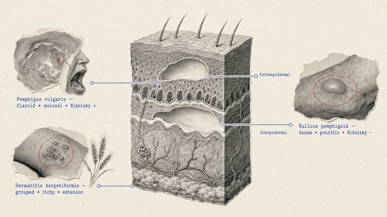
Pemphigus Vulgaris vs Bullous Pemphigoid
This comparison appears in virtually every NEET PG exam. Every row is exam-relevant.
| Feature | Pemphigus Vulgaris | Bullous Pemphigoid |
|---|---|---|
| Age of onset | Middle-aged (40–60 years) | Elderly (over 60) |
| Blister level | Intraepidermal (suprabasal) | Subepidermal |
| Nikolsky sign | Positive | Negative |
| Bulla | Flaccid, ruptures easily | Tense, thick-roofed, does not rupture easily |
| Mucosal involvement | Always involved — oral lesions often first | Rare |
| Acantholysis | Yes — loss of intercellular adhesion | No |
| Target antigen | Desmoglein 3 (oral) and desmoglein 1 (skin) | BP180 and BP230 (hemidesmosomes) |
| Antibody | IgG against desmoglein 3/1 | IgG against BP180 (collagen XVII) and BP230 |
| Direct IF pattern | Net-like, fishnet or chicken-wire | Linear IgG and C3 at the BMZ |
| Indirect IF | IgG on epithelial cell surfaces | IgG along the basement membrane zone |
| Histology | Intraepidermal blister, acantholytic (Tzanck) cells, tombstone row of basal cells | Subepidermal blister with eosinophilic infiltrate |
| Prognosis | More severe, higher mortality | Better |
| Treatment | High-dose systemic steroids plus steroid-sparing agents (azathioprine, rituximab) | Topical or systemic steroids; doxycycline with niacinamide |
Memory pearls: PV — Positive Nikolsky, Pemphigus, intra-epidermal. BP — Basement membrane, Bullous Pemphigoid, subepidermal, Tense blister. IF pattern: PV is net or chicken-wire (intercellular); BP is linear (at the basement membrane).
Pemphigus Histology
On light microscopy: a suprabasal split forming just above the basal layer; acantholytic cells — rounded keratinocytes that have lost cell-to-cell adhesion; a "tombstone" appearance where basal cells remain attached to the basement membrane in a row; and acantholytic cells on Tzanck smear, which is also positive in herpes infections.
On immunofluorescence: direct IF shows IgG deposits in a net-like, chicken-wire pattern on keratinocyte cell surfaces throughout the epidermis, with C3 deposited alongside. Indirect IF detects patient IgG against epidermal cell surfaces.
Bullous Pemphigoid Histology
On light microscopy: a subepidermal blister forming below the entire epidermis; an eosinophil-rich infiltrate in the blister cavity and dermis; no acantholysis, with the epidermis intact and basal cells in the blister roof.
On immunofluorescence: direct IF shows linear IgG and C3 along the basement membrane zone — the linearity is what distinguishes it from PV's net-like pattern. On salt-split skin, the IgG sits on the epidermal side of the split.
Drug-Induced Bullous Pemphigoid
| Drug Class | Examples |
|---|---|
| Diuretics | Furosemide, spironolactone |
| Antihypertensives | Nifedipine, captopril, enalapril |
| Antibiotics | Amoxicillin, ciprofloxacin |
| Antidiabetics | Gliptins (DPP-4 inhibitors) — strong association |
| Antipsychotics | Chlorpromazine |
| Biologics | TNF-alpha inhibitors |
PYQ pattern: the drug class most prominently associated with bullous pemphigoid in recent questions is the DPP-4 inhibitors (gliptins).
Other Bullous Disorders
| Condition | Key Feature | Target Antigen |
|---|---|---|
| Pemphigus foliaceus | Superficial blistering, no mucosal involvement; endemic form in Brazil (fogo selvagem) | Desmoglein 1 only |
| Dermatitis herpetiformis | Intensely pruritic vesicles on elbows, knees, buttocks; associated with coeliac disease | Epidermal transglutaminase |
| Linear IgA disease | Linear IgA at BMZ; "string of pearls" in children | IgA against BP180 |
| Epidermolysis bullosa acquisita | Mechanobullous, at trauma sites; below lamina densa | Collagen VII |
| Porphyria cutanea tarda | Sun-exposed areas, fragile skin, milia; urine fluorescence | Uroporphyrinogen decarboxylase deficiency |
Lichen Planus
Clinical Features — the 4 Ps
Lichen planus is described by the classic four Ps: Pruritic, Purple, Planar (flat-topped) and Polygonal — plus Wickham striae, the whitish lacy lines on the lesion surface.
Sites are the flexor aspects of wrists and forearms, ankles, lower legs and mucosae. Mucosal involvement occurs in 50 to 70 percent of patients, most often as a reticular whitish pattern on the buccal mucosa; the erosive oral form is pre-malignant.
Nail changes include pterygium unguis — adhesion of the proximal nail fold to the nail plate, which is pathognomonic — along with longitudinal ridging and trachyonychia. Koebner phenomenon is positive.
Histology
| Layer | Finding |
|---|---|
| Epidermis | Hyperkeratosis, focal hypergranulosis, irregular acanthosis with saw-tooth rete ridges |
| Dermoepidermal junction | Vacuolar degeneration of basal cells; colloid (Civatte) bodies |
| Dermis | Band-like lichenoid lymphocytic infiltrate hugging the DEJ |
Saw-tooth rete ridges plus a band-like infiltrate is the classic LP pattern. Colloid bodies — also called Civatte or cytoid bodies — are apoptotic keratinocytes appearing as eosinophilic globular structures at the DEJ.
Variants
| Variant | Site or Feature |
|---|---|
| Classic (papular) | Flexor wrists and forearms — most common |
| Oral LP | Buccal mucosa, reticular pattern; erosive form pre-malignant |
| Lichen planus pigmentosus | See below |
| Hypertrophic LP | Thick plaques on legs — the most pruritic variant |
| Bullous LP | Blisters within LP lesions |
| Actinic LP | Sun-exposed areas; commoner in the Middle East and India |
| Lichen planopilaris | Follicular LP causing scarring alopecia |
| Inverse LP | Flexural areas, groin, axillae |
| Ulcerative LP | Chronic painful ulcers on the soles |
Lichen Planus Pigmentosus
Lichen planus pigmentosus is a variant characterised by hyperpigmented macular lesions on sun-exposed and flexural sites, without the classic papular morphology. It is far more common in South Asian and Middle Eastern populations than in Western ones, which makes it disproportionately high-yield for Indian exams.
| Feature | Detail |
|---|---|
| Lesion type | Dark brown to black macules and patches |
| Distribution | Face (especially forehead and temples), neck, flexural folds |
| Sun exposure | Worse on sun-exposed areas — photodistributed |
| Pruritus | Mild to moderate; less intense than classic LP |
| Classic LP features | Usually absent — no Wickham striae, no flat-topped papules |
| Mucosal involvement | Uncommon |
| Course | Chronic; pigmentation may persist long after active disease resolves |
Histology shows vacuolar degeneration of the basal layer and melanin incontinence — melanin within dermal macrophages (melanophages) — with a sparse lymphocytic infiltrate at the DEJ, less band-like than classic LP.
Treatment starts with mandatory sun protection, since UV exposure worsens the pigmentation. Topical corticosteroids of moderate potency are used for limited disease, and topical tacrolimus is the steroid-sparing option preferred on the face. Hydroxychloroquine or low-dose systemic steroids are reserved for resistant cases, and Q-switched Nd:YAG laser can address persistent pigmentation once the disease is inactive.
PYQ pattern: questions ask about the distribution (flexural and sun-exposed), the histological finding (melanin incontinence with melanophages), and the treatment.
Leprosy
Ridley-Jopling Classification
Based on the immunological status of the host — the standard classification tested in NEET PG.
| Type | Bacteria | Lepromin Test | Histology | Clinical Features |
|---|---|---|---|---|
| Tuberculoid (TT) | None | Strongly positive | Well-formed granulomas surrounded by lymphocytes | 1–3 hypopigmented, anaesthetic, dry patches with well-defined borders; prominent nerve enlargement |
| Borderline tuberculoid (BT) | Few | Positive | Less organised granulomas | More lesions than TT, less clear borders |
| Mid-borderline (BB) | Moderate | Equivocal | Mixed picture | Multiple asymmetric lesions, punched-out appearance |
| Borderline lepromatous (BL) | Many | Negative | Poor granulomas, foamy macrophages | Multiple lesions, nerve damage |
| Lepromatous (LL) | Very high | Strongly negative | Foamy macrophages (Virchow cells), no granulomas, globi of organisms | Diffuse thickening, madarosis, ear lobe thickening, leonine facies; nerve damage late |
| Indeterminate (I) | None to few | Variable | Non-specific | Early stage; may progress or regress |
The lepromin test measures delayed hypersensitivity to Mycobacterium leprae antigens. It is positive in tuberculoid disease (good cell-mediated immunity) and negative in lepromatous disease (anergic). It is not diagnostic — it indicates immune status.
WHO Classification for Treatment
| Classification | Lesions | Skin Smear | Treatment |
|---|---|---|---|
| Paucibacillary (PB) | 1–5 patches | Negative | Rifampicin + dapsone for 6 months |
| Multibacillary (MB) | More than 5 patches | Positive | Rifampicin + dapsone + clofazimine for 12 months |
Anti-Leprosy Drugs
| Drug | Mechanism | Key Property |
|---|---|---|
| Rifampicin | RNA polymerase inhibitor | Bactericidal; most potent single agent; monthly supervised dose |
| Dapsone | Dihydropteroate synthase inhibition | Bacteriostatic; daily self-administered; causes haemolytic anaemia and methaemoglobinaemia |
| Clofazimine | DNA binding plus anti-inflammatory | Bacteriostatic and anti-inflammatory (used in Type 2 reactions); causes orange-red skin discolouration |
Leprosy Reactions
| Feature | Type 1 (Reversal) | Type 2 (ENL) |
|---|---|---|
| Mechanism | Delayed hypersensitivity (Type IV) — upregulated CMI | Immune complex deposition (Type III) |
| Spectrum | Borderline types (BT, BB, BL) | Lepromatous (LL, BL) |
| Skin lesions | Existing patches become red, oedematous, tender | New painful red nodules |
| Nerve involvement | Acute nerve function impairment — common | Less prominent |
| Systemic features | Minimal | Fever, malaise, iridocyclitis, orchitis, lymphadenopathy |
| Treatment | Systemic corticosteroids | Thalidomide first-line; systemic steroids |
High-yield: thalidomide is the drug of choice for ENL. Steroids treat Type 1.
Psoriasis
Psoriasis presents as an erythematous plaque with silvery-white scale on extensor surfaces — elbows, knees, scalp, lumbar area and nails. The candle grease sign describes scales detaching in layers like scraping a candle; the Auspitz sign is pinpoint bleeding after scale removal; Koebner is positive.
Nail Changes
| Change | Description |
|---|---|
| Pitting | Most common nail change — small punctate depressions |
| Oil drop sign | Translucent yellow-brown spot under the nail |
| Onycholysis | Separation of nail from nail bed |
| Subungual hyperkeratosis | Thickening under the nail |
| Leukonychia | White discolouration |
Histology
| Finding | Significance |
|---|---|
| Munro's microabscesses | Neutrophil collections in the stratum corneum — pathognomonic |
| Kogoj's spongiform pustule | Neutrophils between keratinocytes in the upper spinous layer |
| Parakeratosis | Retained nuclei in the stratum corneum |
| Regular acanthosis | Elongated rete ridges, test-tube and club shaped |
| Thin suprapapillary plate | Explains the Auspitz sign |
| Dilated tortuous capillaries | In the dermal papillae |
Treatment Ladder
Topical therapy uses corticosteroids first-line for mild disease, with calcipotriol (a vitamin D analogue) — the steroid-calcipotriol combination is standard — plus coal tar, dithranol and tazarotene.
Phototherapy uses narrowband UVB first-line, with PUVA more effective but carrying a higher cancer risk.
Systemic options are methotrexate (most widely used for moderate to severe disease), cyclosporine for rapid control of severe flares, and acitretin for pustular and erythrodermic psoriasis.
| Biologic | Target | Class |
|---|---|---|
| Adalimumab, etanercept, infliximab | TNF-alpha | Anti-TNF |
| Ustekinumab | IL-12 and IL-23 | Anti-IL-12/23 |
| Secukinumab, ixekizumab | IL-17A | Anti-IL-17 |
| Guselkumab, risankizumab | IL-23 (p19 subunit) | Anti-IL-23 |
PYQ pattern: the drug targeting IL-17 is secukinumab; the drug for pustular psoriasis is acitretin.
Vitiligo
| Feature | Detail |
|---|---|
| Pathogenesis | Autoimmune destruction of melanocytes |
| Lesion | Complete depigmentation — milk-white macules with normal skin texture |
| Wood's lamp | Enhanced chalk-white fluorescence |
| Koebner phenomenon | Positive |
| Reverse Koebner | Positive — trauma can cause repigmentation |
| Dermoscopy | Perifollicular pigmentation — a good prognostic sign, indicating a melanocyte reservoir in the follicles |
| Type | Features |
|---|---|
| Generalised (non-segmental) | Most common; bilateral and symmetric; associated with thyroid disease, type 1 diabetes and Addison's |
| Segmental | Unilateral, follows Blaschko's lines, early onset; responds better to grafting |
| Focal | Single or few macules in one body segment |
| Acrofacial | Distal digits and periorificial face |
| Universal | Over 80 percent body involvement — worst prognosis |
Treatment uses topical corticosteroids first-line for limited disease, with topical tacrolimus or pimecrolimus preferred on the face and in children. Narrowband UVB is the most effective phototherapy, with PUVA for generalised disease and excimer laser for localised stable patches. Surgical options — split-thickness grafting and melanocyte transplantation — apply only to stable vitiligo. Oral mini-pulse steroids can arrest progression in active disease.
Scabies
| Feature | Detail |
|---|---|
| Organism | Sarcoptes scabiei var. hominis |
| Pathognomonic lesion | Burrow — a 5–15 mm curved thread-like channel |
| Sites in adults | Finger web spaces, wrists, penis |
| Sites in infants | Palms, soles, face |
| Symptom | Intense pruritus, worse at night |
| Diagnosis | Dermoscopy shows the "jet with contrail" sign — mite plus burrow; microscopy of skin scraping |
| Drug | Notes |
|---|---|
| Permethrin 5% cream | First-line; head to toe, left 8–12 hours, repeated after a week |
| Oral ivermectin | 200 mcg/kg, two doses two weeks apart; first-line for crusted scabies |
| Benzyl benzoate 25% | Traditional, effective, irritating |
| Lindane | Avoid in infants and pregnancy — neurotoxicity |
| Sulfur ointment | Safest in infants and pregnancy |
Norwegian or crusted scabies is a hyperinfestation seen in immunocompromised patients — HIV, the elderly, long-term corticosteroid users — with millions of mites, high contagiousness and crusted hyperkeratotic plaques. Treatment combines ivermectin with a topical scabicide.
Drug-Induced Skin Reactions
| Reaction | Drug | Key Feature |
|---|---|---|
| Fixed drug eruption | Tetracyclines, NSAIDs, cotrimoxazole | Recurs at the same site on re-exposure |
| Stevens-Johnson syndrome | Sulfonamides, carbamazepine, allopurinol, lamotrigine | Under 10 percent BSA, mucosal involvement |
| Toxic epidermal necrolysis | Same drugs, more severe | Over 30 percent BSA; Nikolsky positive |
| DRESS syndrome | Anticonvulsants, allopurinol, sulfonamides | Eosinophilia with organ involvement; onset 2–8 weeks |
| Photosensitivity | Tetracyclines, amiodarone, chlorpromazine, fluoroquinolones | Sun-exposed distribution |
| Lichenoid drug reaction | Antimalarials, gold, captopril, thiazides | Mimics lichen planus |
| Acneiform eruption | Steroids, isoniazid, lithium, OCP | Monomorphic comedones |
| Alopecia | Cytotoxics, heparin, anticoagulants, retinoids | Diffuse hair loss |
Wood's Lamp Findings
| Organism or Condition | Colour |
|---|---|
| Microsporum canis (tinea capitis) | Green to yellow-green |
| Microsporum audouinii | Brilliant green |
| Trichophyton schoenleinii (favus) | Pale greenish-white |
| Pityriasis versicolor (Malassezia) | Yellow-green, coppery-orange |
| Pseudomonas aeruginosa | Green |
| Erythrasma (Corynebacterium minutissimum) | Coral red |
| Porphyria cutanea tarda | Pink-red urine fluorescence |
| Vitiligo | Chalk-white enhancement |
PYQ pattern: erythrasma fluoresces coral red because Corynebacterium produces porphyrins; Microsporum tinea capitis fluoresces green to yellow-green.
High-Yield Dermatology PYQ Checklist
Work through this alongside a timed NEET PG HAMMER test to see which items actually hold up under exam conditions.
- Nikolsky sign — positive in PV, negative in BP
- Koebner phenomenon — psoriasis, LP, vitiligo, not pemphigus
- Reverse Koebner — vitiligo
- Pseudo-Koebner — viral warts, molluscum
- Auspitz sign — psoriasis; mechanism is suprapapillary thinning
- PV target antigen — desmoglein 3 (oral), desmoglein 1 (skin)
- BP target antigen — BP180 (collagen XVII), BP230
- PV immunofluorescence — net or chicken-wire, intercellular
- BP immunofluorescence — linear at the BMZ
- Colloid (Civatte) bodies — lichen planus
- Saw-tooth rete ridges with band-like infiltrate — lichen planus
- Munro's microabscesses — psoriasis, pathognomonic
- Kogoj's spongiform pustule — psoriasis
- Lichen planus pigmentosus — melanin incontinence; flexural and sun-exposed
- Ridley-Jopling TT — lepromin strongly positive, no AFB, well-formed granulomas
- Ridley-Jopling LL — lepromin negative, very high AFB, Virchow cells
- WHO paucibacillary — 5 or fewer lesions; rifampicin and dapsone for 6 months
- WHO multibacillary — more than 5 lesions; add clofazimine, 12 months
- Type 1 leprosy reaction — borderline types; steroids
- Type 2 leprosy reaction (ENL) — LL and BL; thalidomide first-line
- Anti-IL-17 for psoriasis — secukinumab, ixekizumab
- Anti-IL-23 for psoriasis — guselkumab, risankizumab
- Permethrin 5% — first-line for scabies
- Norwegian scabies — immunocompromised; ivermectin
- Erythrasma on Wood's lamp — coral red
- Microsporum tinea capitis on Wood's lamp — green to yellow-green
- Dermoscopy in vitiligo — perifollicular pigmentation indicates good prognosis
- Pterygium unguis — nail lichen planus, pathognomonic
Practise this on Reflex
Turn what you just read into recall with 14 years of tagged PYQs.
FAQ
Frequently asked questions
The questions aspirants ask most about this topic.
Pemphigus vulgaris versus bullous pemphigoid — comparison, histology and immunofluorescence — plus the Koebner family of phenomena, the Auspitz sign, Ridley-Jopling leprosy classification, leprosy reactions and lichen planus clinical features appear in nearly every cycle. These seven clusters cover roughly 60 to 70 percent of Dermatology marks.
The Nikolsky sign is positive when lateral pressure on apparently normal skin causes it to slide off, indicating poor epidermal adhesion. It is positive in pemphigus vulgaris, where the split is intraepidermal and suprabasal, and negative in bullous pemphigoid, where the blister is subepidermal and tense. It is also positive in staphylococcal scalded skin syndrome and toxic epidermal necrolysis.
It is a variant presenting as dark brown to black macules on sun-exposed areas and flexural folds, particularly common in South Asian patients. Unlike classic LP it has no flat-topped papules and no Wickham striae. Histology shows melanin incontinence with melanophages in the dermis and vacuolar basal degeneration with a sparse infiltrate. Treatment is strict sun protection, topical corticosteroids and topical tacrolimus.
Type 1, the reversal reaction, occurs in borderline types and is a delayed hypersensitivity reaction — existing patches inflame and acute nerve damage may follow. It is treated with systemic corticosteroids. Type 2, erythema nodosum leprosum, occurs in lepromatous disease through immune complex deposition, producing new painful nodules with fever and iridocyclitis. Thalidomide is first-line.
For mild disease, topical corticosteroids combined with calcipotriol. For moderate to severe disease, methotrexate or cyclosporine systemically, or narrowband UVB phototherapy. Biologics targeting TNF-alpha (adalimumab), IL-17 (secukinumab) or IL-23 (guselkumab) are used where conventional systemic therapy fails, and these drug-target pairings are increasingly tested.
Strict photoprotection with broad-spectrum sunscreen is mandatory, since UV exposure worsens the pigmentation. Mid-potency topical corticosteroids reduce inflammation, and topical tacrolimus 0.1% is preferred on the face to avoid steroid side effects. Hydroxychloroquine is used for widespread or resistant disease, and Q-switched Nd:YAG laser helps with persistent pigmentation once the disease is inactive.
About the author
Stay ahead in your preparation
Get expert tips, exam updates, and high-yield insights delivered straight to your inbox.
No spam. Unsubscribe anytime.
More in Subject Notes
View all → Dermatology
Pityriasis Rosea for NEET PG: The Herald Patch, the Christmas-Tree Pattern and the Papulosquamous Differential
 Surgery
Burns for NEET PG: The Rule of Nines, Burn Depth and the Parkland Formula Worked Step by Step
 Radiology
FAST Scan for NEET PG: The Four Windows, eFAST, and What a Positive Result Means
 Psychiatry