Intussusception for NEET PG: The Classic Triad, Target Sign and Reduction Methods
Reflex · 8 Nov 2026 · 10 min read
Last updated: 8 Nov 2026
On this page
A previously well infant screams, draws up the legs, settles, and then screams again. Between episodes the child looks almost normal, which is exactly why intussusception is missed. This post walks through the condition the way a clinical stem does: who gets it, what the features are, which imaging sign confirms it, and how it is reduced.
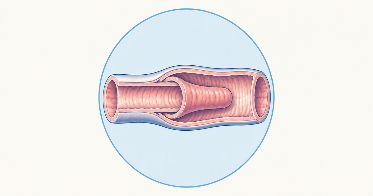
Intussusception means one segment of bowel telescopes into the segment just distal to it. The inner part is the intussusceptum and the outer part is the intussuscipiens. The result is obstruction, then venous congestion, then ischaemia if the telescoped bowel is not released.
Key Takeaways
Boys are affected more than girls, and the age band to remember is 3 months to 2 years, with the peak at 4 to 9 months.
Fewer than 15% of children have all three parts of the classic triad (colicky pain, palpable mass, red currant jelly stool), so do not wait for all three.
Red currant jelly stool is a late sign that points towards ischaemia.
Ultrasound is the first-line investigation: target (doughnut) sign on transverse view, pseudokidney sign on longitudinal view.
Air or contrast enema reduction is the first-line treatment in a stable child without peritonitis, shock or perforation.
Surgery is for failed reduction, peritonitis or perforation, suspected necrosis, recurrent episodes or an organic lead point.
What Is Intussusception and Who Gets It?
According to the Royal Children's Hospital Melbourne guideline, the peak is at 4 to 9 months, and the condition is most common between 3 months and 2 years of age. Children account for about 95% of cases, although intussusception can occur at any age (Radiopaedia). A 2026 Frontiers in Pediatrics review reports a male to female ratio of about 2 to 3 to 1.
Most childhood cases have no identifiable cause. Over 95% are primary (idiopathic) in the same review, and Radiopaedia puts the proportion with no lead point at about 90% of children.
Site: why ileocolic is the default answer
The ileocolic type is the commonest in children. Radiopaedia gives about 75 to 95% of paediatric cases, and a Turkish series of 183 children found 164 (89.6%) were ileocolic (Dorterler and Kocaman, Cureus 2019). When a stem simply says "intussusception in an infant", think ileocolic first.
Small-bowel (ileoileal) intussusception is the exception to learn. In IgA vasculitis (Henoch-Schonlein purpura), intussusception is typically ileoileal (Radiopaedia).
Why It Happens: Primary Versus Pathological Lead Points
In infants, the usual story is a viral trigger. Adenovirus or rotavirus infection causes reactive lymphoid hyperplasia of Peyer's patches, and the thickened ileocaecal region acts as the lead point. Peristalsis then pushes the proximal bowel into the distal lumen. A mobile infant mesentery and the difference in calibre between ileum and caecum help the process along (Front Pediatr 2026).
A pathological lead point is found in only about 6 to 10% of children. In the Cureus series, 14 of 183 children (7.6%) had one, and mesenteric lymphadenitis was the commonest.
The table below separates the two situations, because the lead point changes the management.
| Feature | Primary (idiopathic) | Pathological lead point |
|---|---|---|
| Share of paediatric cases | Over 95% (Front Pediatr 2026) | About 6 to 10% (Cureus 2019) |
| Typical age | 6 months to 2 years | Older children more likely |
| Mechanism | Lymphoid hyperplasia after viral infection | Meckel diverticulum, polyps (for example Peutz-Jeghers), lymphoma, recent bowel surgery |
| Usual treatment | Enema reduction | Organic lesion is an indication for surgery |
Sources: Front Pediatr 2026, Cureus 2019 and the RCH guideline.
In adults the picture reverses. Radiopaedia reports a lead point in up to 90% of adult cases, with malignancy in about 65%. That is why adults are not managed like infants.
Rotavirus vaccine and intussusception
The vaccine link needs careful wording. The WHO Global Advisory Committee on Vaccine Safety reported a small potential risk, in the range of 1 to 2 cases per 100,000 first doses, mainly within 7 days of dose 1. WHO concluded that the benefit of vaccination outweighed the risk (WHO GACVS).
Indian surveillance of 589 infants at 27 hospitals looked at Rotavac dose 1. The relative incidence was 0.83 for days 1 to 7 and 0.35 for days 8 to 21, but both confidence intervals were wide (0.0 to 3.00 and 0.0 to 1.09), each from only 2 cases. The authors concluded that Rotavac was not associated with intussusception (NEJM 2020).
For the doses and ages themselves, see the immunization schedule post or practise with the paediatric immunization questions.
The Classic Triad and Other Clinical Features
Red currant jelly stool, a palpable abdominal mass and intermittent colicky abdominal pain make up the classic triad. The important point is that the full triad is present in fewer than 15% of children (RCH guideline). A child can have intussusception with only one or two of these features.
Each part of the triad
- Colicky pain. Quiet periods separate the episodes, in which severe pain makes the child draw up the legs. Between episodes the child may seem well or lethargic.
- Sausage-shaped mass. Typically in the right abdomen, though it may cross the midline into the epigastrium. It is best felt when the child is settled between episodes (RCH). The Frontiers review also describes an empty right lower abdomen on palpation.
- Red currant jelly stool. Mucus and blood are passed together in the stool. It is a late sign that suggests ischaemia (RCH). The Frontiers review describes dark brown bloody stool about 6 to 12 hours after onset, from mucosal ischaemic necrosis.
The memory aid here is simple: the stool tells you about time. If the stem already shows red currant jelly stool, the bowel has probably been compromised for hours, and the child is further along the pathway than the pain alone suggests.
Atypical presentations
Not every infant reads the textbook. Lethargy, feed refusal, pallor and vomiting can be the main features, and bilious vomiting suggests obstruction (Front Pediatr 2026). In an infant with unexplained lethargy and pallor, keep intussusception on the list.
Imaging: Ultrasound First, Signs to Recognise
Ultrasound is the first-line investigation. The RCH guideline reports sensitivity and specificity both above 98% with an experienced paediatric sonographer, while the Frontiers review quotes sensitivity up to 96% and specificity of 92%. A fair summary is about 92 to 98%, depending on operator and setting.
A negative bedside scan must not be used to exclude the diagnosis, even though the RCH guideline gives point-of-care ultrasound a sensitivity of 95% and a specificity of 98%.
Ultrasound signs
| Sign | View | What you see |
|---|---|---|
| Target (doughnut) sign | Transverse | Concentric rings of the telescoped bowel |
| Pseudokidney sign | Longitudinal | Layered bowel resembling a kidney |
| Absent blood flow in the intussusceptum | Doppler | Contraindication to enema reduction (Radiopaedia) |
The Frontiers review also uses the names concentric circle sign and tube sign (longitudinal view) for the same appearances. Learn target and pseudokidney as a pair: same lesion, cut in two planes. The Doppler row follows Radiopaedia.
Plain X-ray and CT
A plain abdominal film has a sensitivity below 50%, so a normal film does not exclude intussusception. The crescent sign is described on plain films (RCH). CT is primarily used for complex cases or when the ultrasound diagnosis is unclear (Front Pediatr 2026).
Reduction Methods: Air and Contrast Enema
Non-operative reduction is the first-line treatment in a stable child. Air or water-soluble contrast is delivered through a rectal catheter, and the procedure is done where paediatric surgery is available (RCH).
Contraindications
Go straight to surgery, not to an enema, when any of these is present:
- Peritonitis.
- Shock or an unstable child.
- Radiological perforation or free air.
- Absent blood flow in the intussusceptum on ultrasound (Radiopaedia).
How well does reduction work?
Success rates vary with the delay before treatment and the setting. RCH quotes more than 80%, with repeat enemas adding about 10%. The Frontiers review says air enema can exceed 95% in early cases and falls when symptoms last more than 24 hours or ischaemia is present. The Cureus series reported 74.7% (115 of 154) for pneumatic reduction. A safe summary is roughly 75 to 95%, depending on duration and setting.
Symptoms for more than 24 hours and ischaemia are the main predictors of failure. The perforation rate in the literature ranges from 0.1 to 3% (Cureus 2019).
Air, contrast or ultrasound-guided saline?
A 2024 PLOS ONE meta-analysis pooled 49 studies, randomized and non-randomized, and the success outcome used 48 of them. Ultrasound-guided hydrostatic reduction beat fluoroscopy-guided air reduction on success (RR 1.128, 95% CI 1.099 to 1.157). It also had less perforation (RR 0.405, 95% CI 0.244 to 0.670) and less recurrence (RR 0.391, 95% CI 0.269 to 0.569) (Liu et al., PLOS ONE 2024).
For revision, the principle matters more than the numbers: the enema is both diagnostic and therapeutic, and it is only safe when the child is stable and the bowel is likely viable.
When Surgery Is Needed
Surgery follows a failed enema or any contraindication to one. The Frontiers review lists these indications:
- Failed reduction.
- Onset beyond 24 to 48 hours with suspected necrosis or perforation.
- Recurrent intussusception (three or more episodes).
- Small-bowel intussusception.
- An organic lesion such as Meckel diverticulum, duplication, polyp or tumour.
Radiopaedia adds peritonitis, rectal blood and protracted symptoms. The operation can be laparoscopic or open. If the bowel is viable and there is no organic lesion, it is reduced manually. If it is necrotic, irreducible or has a lesion, the segment is resected and an anastomosis is made (Frontiers review).
Recurrence After Reduction
Recurrence is reported at about 6 to 15% depending on the series. The Cureus paper cites 6 to 10% after hydrostatic or pneumatic reduction and 10% after surgical reduction, while Radiopaedia gives 9 to 15%. Most recurrences happen within 24 to 48 hours (RCH). Three or more episodes in the same child points to a lead point and is an indication for surgery.
Common Mistakes When Revising Intussusception
- Waiting for the full triad. It is present in fewer than 15% of children, so the diagnosis rests on the pattern of episodic pain, lethargy or vomiting plus imaging.
- Treating red currant jelly stool as an early sign. It is late and suggests ischaemia.
- Trusting a normal X-ray. Sensitivity is below 50%.
- Attempting an enema in a child with peritonitis, shock or free air. These go to surgery.
- Mixing up the vaccine data. Say "small potential risk, mainly in the first week after dose 1, benefit outweighs risk", not that the vaccine causes it.
- Quoting the wrong age band. Use 3 months to 2 years with a peak at 4 to 9 months.
Quick Revision Sequence
If a stem gives you an infant with episodic screaming, think of intussusception. Then run through the sequence:
- Confirm with ultrasound (target or pseudokidney sign).
- Check for peritonitis, shock, free air or absent blood flow.
- If none is present, reduce with an air or contrast enema.
- Operate if reduction fails or any contraindication is present.
- Watch for recurrence in the first 24 to 48 hours.
Conclusion
Intussusception is a pattern-recognition topic. The age band (3 months to 2 years), the episodic pain, the late appearance of red currant jelly stool and the target and pseudokidney signs on ultrasound make up the diagnosis. The treatment is a stable-child decision: enema reduction when it is safe, surgery when it is not or when it fails. To test yourself, try the paediatric gastroenterology practice questions.
Practise this on Reflex
Turn what you just read into recall with 14 years of tagged PYQs.
FAQ
Frequently asked questions
The questions aspirants ask most about this topic.
Red currant jelly stool, a palpable sausage-shaped abdominal mass and intermittent colicky abdominal pain form the classic triad. Fewer than 15% of children show all three features, so the diagnosis should not wait for them.
Children between 3 months and 2 years are most often affected, with the peak at 4 to 9 months. Boys are affected more often than girls, although intussusception can occur at any age.
On ultrasound, look for the target (doughnut) sign on the transverse view and the pseudokidney sign on the longitudinal view. Absent blood flow in the intussusceptum on Doppler is a contraindication to enema reduction.
Enema reduction is contraindicated in peritonitis, shock or an unstable child, and radiological perforation or free air. Absent blood flow in the intussusceptum on ultrasound is also a contraindication. These children go to surgery.
Surgery is indicated after failed enema reduction, when symptoms began more than 24 to 48 hours earlier with suspected necrosis or perforation, with recurrent episodes (three or more), in small-bowel intussusception, or when an organic lesion such as a Meckel diverticulum or polyp is the lead point.
WHO reports a small potential risk (in the range of 1 to 2 cases per 100,000 first doses), particularly within the first 7 days after dose 1, and concluded that the benefit of vaccination outweighed the risk. Indian surveillance of Rotavac found no association with intussusception, although the dose-1 confidence intervals were wide.
Stay ahead in your preparation
Get expert tips, exam updates, and high-yield insights delivered straight to your inbox.
No spam. Unsubscribe anytime.
More in Pediatrics
View all → Pediatrics
Neonatal Jaundice for NEET PG: Physiological vs Pathological Criteria and the Phototherapy Logic
A revision guide to neonatal jaundice: how to separate physiological from pathological jaundice, how phototherapy and exchange decisions are structured, and what kernicterus and cholestasis look like.
 Pediatrics
Kawasaki Disease for NEET PG: The Five Criteria, the 10-Day IVIG Window and the Coronary Risk
What counts as Kawasaki disease, why the 10-day IVIG window matters, how aspirin is used, and how coronary artery involvement is graded and followed.
 Pediatrics
Tetralogy of Fallot for NEET PG: One Septal Shift, Four Defects, and the Tet Spell
One shifted septum explains the four defects of Tetralogy of Fallot, the boot-shaped heart, the fading murmur and the stepwise management of a tet spell.
 Pediatrics