Tetralogy of Fallot for NEET PG: One Septal Shift, Four Defects, and the Tet Spell
By Dr. Utsav Bhattacherjee, MBBS, MBA · 23 Oct 2026 · 18 min read
Last updated: 23 Oct 2026
On this page
Tetralogy of Fallot (TOF) is the most common cyanotic congenital heart defect overall, although transposition of the great arteries is the lesion that most often presents with cyanosis in the newborn period. One structural change explains almost everything a question can ask about TOF: the blue baby, the ejection murmur, the boot-shaped heart, the squatting toddler and the sudden spell. Learn that single abnormality first and the four-part definition, the investigations and the spell management stop being separate lists to memorize. This post builds the logic in that order, then covers the bedside findings, the imaging traps and the treatment ladder.
What Tetralogy of Fallot Is: One Shift, Four Defects
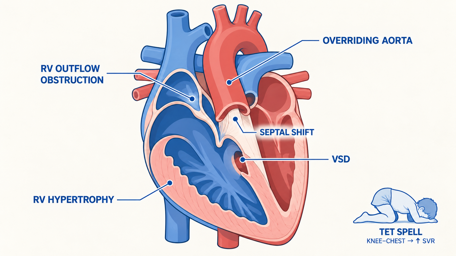
The root problem is a displacement of the infundibular (outlet) septum. In TOF it sits too far forward and upward, which is described as an anterosuperior deviation. The standard explanation is that this one shift does two things at once. It malaligns the septum against the rest of the ventricular septum, leaving a large ventricular septal defect (VSD), and it narrows the path from the right ventricle to the lungs, which is right ventricular outflow tract (RVOT) obstruction.
The aorta then sits over the defect, so it overrides the septum, and the right ventricle thickens because it pumps against the obstruction. That gives the four components: pulmonary stenosis, a large VSD, an overriding aorta and right ventricular hypertrophy (RVH). A common memory aid is PROV: Pulmonary stenosis, Right ventricular hypertrophy, Overriding aorta, VSD.
| Component | What it is | What it does |
|---|---|---|
| Pulmonary stenosis | Narrowing at the infundibulum, the pulmonary valve, or both | Sets how much blood reaches the lungs and produces the murmur |
| Ventricular septal defect | Large defect, malaligned toward the front | Lets deoxygenated blood cross to the aorta; usually silent because it is large |
| Overriding aorta | Aorta positioned over the defect | Receives blood from both ventricles |
| Right ventricular hypertrophy | Thickened right ventricular wall | A result of pumping against the obstruction; gives the upturned apex and right axis deviation |
Only two of the four decide the physiology. The VSD gives blood a path from the right ventricle to the aorta, and the obstruction decides how much blood takes that path instead of going to the lungs. The hypertrophy is a consequence, and the override simply lines the aorta up with the defect.
Estimates put TOF at about 5% to 10% of congenital heart disease. Roughly 15% to 25% of cases occur with a syndrome or chromosomal abnormality, most often Down syndrome or the 22q11.2 deletion that causes DiGeorge syndrome. A right aortic arch is present in about 25% of patients. Surgeons also look for a coronary artery crossing the outflow tract, such as the left anterior descending artery arising from the right coronary artery, because it changes how the outflow can be opened.
Why the Baby Turns Blue: Two Resistances Decide the Shunt
The VSD in TOF is large and does not restrict flow, so the direction of the shunt depends on the resistance on either side of it. On one side is the obstruction in the RVOT. On the other is the systemic vascular resistance (SVR) that the left ventricle pumps against. When the RVOT obstruction exceeds SVR, deoxygenated blood takes the easier route across the VSD into the aorta, and the child is cyanotic.
When the obstruction is mild, the shunt runs mostly left to right and the child stays pink. That produces a spectrum worth knowing:
- Pink tet: mild obstruction. There is little or no cyanosis, and the child may come to attention only because of a loud murmur. With very mild obstruction the picture can resemble a large VSD, with extra blood flowing to the lungs.
- Classic TOF: moderate to severe obstruction, with cyanosis, hypercyanotic spells and squatting.
- TOF with pulmonary atresia: the extreme end. Lung blood flow comes through the ductus arteriosus, aortopulmonary collaterals, or both, so the newborn can be severely cyanotic, and prostaglandin E1 is used when the lungs depend on the duct.
Picture an illustrative case: a 4-month-old brought for poor feeding. His mother has noticed a bluish tinge around his lips when he cries, and he tires halfway through a feed. He has central cyanosis, a harsh systolic murmur along the left sternal border and a single second heart sound. His saturation barely rises with high-concentration oxygen, which is the response expected with a fixed right-to-left shunt rather than a simple lung problem.
Bedside Findings: What the Examination Shows
Cyanosis may be present from birth in severe obstruction or pulmonary atresia, while other babies are pink and are found because of a murmur. Cyanosis worsens with feeding and crying. Chronic hypoxemia leads to polycythemia and clubbing, and the clubbing causes post covers the grades and Schamroth's window test.
Start with the murmur, because it is where the logic matters. It is a harsh systolic ejection murmur, typically grade 3 to 5 out of 6, heard best at the left mid to upper sternal border, and it comes from the pulmonary stenosis. The VSD is large and usually silent. The second heart sound is single because the pulmonary component is soft or inaudible, and a systolic thrill may be palpable.
The murmur's intensity runs opposite to severity. As the obstruction worsens, less blood crosses the outflow tract and the murmur gets softer, and during a spell it fades or disappears altogether. That reverses the instinct that a louder murmur means a worse lesion, a trap the murmur grading post explains in general terms. A few references state the opposite relationship, so anchor on the flow logic: the murmur tracks blood crossing the outflow tract, and that flow falls as the obstruction tightens.
Toddlers and older children often squat spontaneously, and infants are placed in the knee-chest position. Squatting raises systemic vascular resistance and aortic pressure, which reduces the right-to-left shunt and raises arterial oxygen saturation.
Hypercyanotic spells peak at 2 to 4 months of age and occur mainly between a few months and 2 years. Typical triggers are crying, feeding, defecation, waking, sudden tachycardia and hypovolemia. During a spell the child becomes deeply blue, limp and irritable, with fast, deep breathing.
Investigations: Boot, Oligemia and Right Axis
The chest X-ray shows a heart of normal size with an upturned apex, from right ventricular hypertrophy, and a concave main pulmonary artery segment, because the pulmonary outflow is small. Together these give the boot-shaped heart. The lung fields look dark, meaning decreased pulmonary vascularity, because blood flow to the lungs is reduced. A right aortic arch, seen in about 25%, is a useful supporting clue.
The ECG shows right axis deviation and right ventricular hypertrophy, with a prominent R wave in lead V1. Echocardiography is the diagnostic test: it defines the VSD, the level and severity of the obstruction, the side of the aortic arch and often the proximal coronary course. CT, MRI or catheterization define the coronary and pulmonary artery anatomy when an anomaly is suspected. At the bedside, a hyperoxia test helps: the child breathes high-concentration oxygen, and a clear rise in arterial oxygen (PaO2) suggests a lung cause, while little or no rise suggests a fixed right-to-left shunt.
| Lesion | Classic chest X-ray sign | Pulmonary vascularity |
|---|---|---|
| Tetralogy of Fallot | Boot-shaped heart | Decreased in classic TOF; can be normal or increased in pink tet |
| Transposition of the great arteries | Egg on a string | Increased |
| Tricuspid atresia | Often a normal or slightly enlarged heart with right atrial enlargement; occasionally mimics TOF | Decreased in the most common form |
| Ebstein anomaly | Box-shaped or balloon-shaped cardiomegaly from right atrial enlargement | Decreased, or normal if the child is not cyanotic |
The ECG axis helps separate TOF from its closest mimic. Tricuspid atresia also has dark lung fields and can occasionally give a boot-shaped heart, but it characteristically shows left axis deviation, between 0 and minus 90 degrees. A cyanotic infant with dark lung fields and a left axis therefore classically points to tricuspid atresia rather than TOF. The pointer is not exclusive: an atrioventricular canal defect can also give a left axis, and the axis is usually not left-deviated when tricuspid atresia coexists with transposition.
A memory aid for the main cyanotic lesions is the five T's: Tetralogy of Fallot, Transposition of the great arteries, Tricuspid atresia, Truncus arteriosus and Total anomalous pulmonary venous connection. It is a memory aid and not a complete list, since Ebstein anomaly and pulmonary atresia also cause cyanosis. The supracardiac form of total anomalous pulmonary venous connection gives the snowman sign on the film.
Managing a Tet Spell: The Logic Behind Each Step
A tet spell is an acute drop in pulmonary blood flow, with a surge of right-to-left shunting across the VSD. The classic explanation is dynamic spasm of the infundibulum on top of the fixed narrowing. A fall in SVR, and a rise in pulmonary vascular resistance from fast breathing, hypoxia and acidosis, also push blood across the VSD, and the exact mechanism is not settled. Anything that raises catecholamines or lowers SVR can set a spell off, including crying, pain, fever, dehydration and a warm environment. Each treatment step therefore does one of three jobs: it raises SVR, it relaxes the infundibulum and lowers pulmonary vascular resistance, or it corrects volume and acidosis.
| Step | What it does |
|---|---|
| Knee-chest position and a calm environment | Raises SVR and cuts systemic venous return; calm lowers the catecholamine surge that drives infundibular spasm |
| Oxygen | Dilates the pulmonary vessels and lowers pulmonary vascular resistance; give it if it does not distress the child |
| IV fluids | Treat hypovolemia, a recognized trigger, and improve right ventricular filling |
| Morphine | Sedates and calms the child, which is thought to reduce the catecholamine-driven spasm |
| Phenylephrine | An alpha agonist that raises SVR, the drug equivalent of the knee-chest position |
| Propranolol or esmolol | Beta-blockers are thought to relax the infundibular spasm, and they slow the heart rate |
| Sodium bicarbonate | Corrects metabolic acidosis when it is present |
| Ketamine | Added when standard measures fail; it sedates and tends to raise SVR |
| Airway control, extracorporeal support or urgent surgery | For refractory spells: intubation with anesthesia and paralysis, ECMO, or an emergency shunt or repair |
The order above follows an immediate-then-escalating sequence, and standard references differ on where the middle drugs and ketamine fall, and on doses. Quoted doses, for IV propranolol in particular, also differ between references, so this post gives the logic and leaves doses to your unit's protocol. What does not change is the opening move: position, calm, oxygen.
Two cautions help in practice. Avoid repeated cannulation attempts that make the baby cry before the spell has settled, because crying prolongs it. And remember that drugs which stimulate beta receptors can tighten the infundibulum, which is why beta-blockers, not beta agonists, belong in the spell algorithm.
Back to the 4-month-old: during a bath he screams, turns deeply blue, goes limp and breathes fast and deep, and the murmur that was loud in the clinic is now barely audible. The first minute is positioning, calming and oxygen, with his knees brought up to his chest in the parent's arms. IV access and drugs follow only if the spell does not settle. If it still does not respond to the standard drugs, the next steps are ketamine and then airway control, extracorporeal support or urgent surgery.
Definitive Treatment: Timing of Repair and the Role of a Shunt
Medical measures only bridge to surgery. Oral propranolol can be used to prevent recurrent spells while the child waits. Complete repair closes the VSD so that the left ventricle ejects into the aorta, and it relieves the right ventricular outflow obstruction.
Elective repair is done in early infancy. Most references quote windows of about 2 to 6 months, depending on the source and the center, and repair is brought forward if the child is symptomatic or the obstruction is severe.
A palliative modified Blalock-Taussig shunt, a small tube from the subclavian artery to the pulmonary artery, is used when complete repair has to wait. Reasons include a small or low-birth-weight neonate, small branch pulmonary arteries, unfavorable coronary anatomy and other major comorbidity. In neonates whose lung blood flow depends on the ductus, prostaglandin E1 keeps it open until the shunt or repair, and catheter-based stenting of the ductus or the outflow tract is a less invasive option in some high-risk infants.
After Repair: Why Follow-Up Never Ends
Repair corrects the anatomy but does not end the story. The main late problem is pulmonary regurgitation. It is very common after repair, close to universal when a patch is placed across the pulmonary valve ring (a transannular patch), and it overloads the right ventricle over decades. Sustained ventricular tachycardia is reported in roughly 5% to 15% of adults with repaired TOF in different series, and sudden cardiac death in roughly 0.1% to 0.2% per year. Pulmonary insufficiency is the most common reason for late reoperation in adults, while early reinterventions are mostly for residual or recurrent pulmonary stenosis.
Figures vary between surgical series, so treat these as orders of magnitude, not exam constants. Endocarditis prophylaxis is recommended before repair, and after repair it continues for the first 6 months, or longer if a residual defect lies next to a patch or prosthetic material.
High-Yield Traps and One-Line Answers
- The murmur is pulmonary stenosis, not the VSD, which is large and usually silent.
- A softer murmur means worse obstruction, and in a spell it fades or disappears.
- Shunt direction depends on RVOT obstruction versus SVR, so squatting, knee-chest and phenylephrine all help by raising SVR.
- Boot-shaped heart, dark lung fields and right axis deviation point to TOF. Dark lung fields with left axis deviation point to tricuspid atresia.
- Egg on a string with increased pulmonary vascularity is transposition, not TOF.
- A right aortic arch occurs in about 25%, and Down syndrome and the 22q11.2 deletion are the syndromes to link with TOF.
- Spells peak at 2 to 4 months, and the first step is the knee-chest position.
- Pink tet looks like a VSD with a murmur and little or no cyanosis.
Pair this with the Kawasaki disease post, which approaches the pediatric heart from the opposite direction: an acquired systemic vasculitis of medium-sized arteries, most significantly the coronary arteries, rather than a structural defect. Then turn the notes into recall with Pediatrics questions in Reflex practice.
Practise this on Reflex
Turn what you just read into recall with 14 years of tagged PYQs.
FAQ
Frequently asked questions
The questions aspirants ask most about this topic.
Pulmonary (right ventricular outflow) stenosis, a large ventricular septal defect, an overriding aorta and right ventricular hypertrophy. All four follow from one anterosuperior shift of the infundibular septum. The memory aid PROV covers them: Pulmonary stenosis, Right ventricular hypertrophy, Overriding aorta, VSD.
Squatting, or the knee-chest position in infants, raises systemic vascular resistance and aortic pressure. That reduces the right-to-left shunt across the VSD, so more blood reaches the lungs and arterial oxygen saturation rises.
Place the child in the knee-chest position and keep them calm, and give oxygen if the child tolerates it. If the spell persists, add IV access with fluids, then morphine, phenylephrine or a beta-blocker such as propranolol or esmolol, with sodium bicarbonate if there is metabolic acidosis. Spells that still do not respond need ketamine, airway support, extracorporeal support or urgent surgery.
The murmur comes from blood crossing the narrowed outflow tract. In a spell the infundibulum tightens and less blood reaches the lungs, so the murmur softens or disappears. A quiet murmur in a blue, distressed child signals a worse spell, not an improvement.
Complete surgical repair, usually done in infancy, closes the VSD and relieves the outflow obstruction. It is better described as corrected than cured, because pulmonary regurgitation, arrhythmia and the need for reintervention can appear decades later, so follow-up is lifelong.
Blue baby is a lay phrase for cyanosis in an infant, and Tetralogy of Fallot is one of the heart defects that causes it. The phrase is also used for methemoglobinemia, so in a clinical stem look at the murmur, the chest X-ray and the arterial blood gas. Pulse oximetry does not separate the two, because the reading stays low with oxygen in both. On the blood gas, methemoglobinemia has a normal PaO2 and chocolate-brown blood, whereas a cyanotic heart defect has a low PaO2.
About the author
Stay ahead in your preparation
Get expert tips, exam updates, and high-yield insights delivered straight to your inbox.
No spam. Unsubscribe anytime.
More in Subject Notes
View all → Pediatrics
Kawasaki Disease for NEET PG: The Five Criteria, the 10-Day IVIG Window and the Coronary Risk
What counts as Kawasaki disease, why the 10-day IVIG window matters, how aspirin is used, and how coronary artery involvement is graded and followed.
 Pediatrics
Failure to Thrive for NEET PG: The Organic vs Non-Organic Branch That Decides the Workup
 Pediatrics
Immunization Schedule for NEET PG: The Complete Age-Wise Breakdown
The National Immunization Schedule by age — birth, 6–14 weeks, 9 months, and the boosters examiners test.
 Microbiology
Malaria Life Cycle for NEET PG: Species, Smear Morphology, Complications and Treatment
How the malaria parasite moves between mosquito and human, what separates the species on a smear, and how diagnosis, complications and treatment follow from the life cycle.