Red Eye for NEET PG: Conjunctivitis, Keratitis, Iritis and Angle-Closure Glaucoma Compared
By Dr. Utsav Bhattacherjee, MBBS, MBA · 28 Sept 2026 · 12 min read
Last updated: 1 Oct 2026
On this page
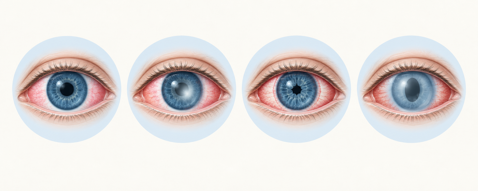
A red eye has a long differential, but almost every exam question on it comes down to telling four conditions apart: conjunctivitis, keratitis, iritis and acute angle-closure glaucoma. Three bedside findings, pain, vision and the pupil, separate them faster than any test. Learn those three, and the differential sorts itself.
Start With Three Questions: Pain, Vision and the Pupil
A painless red eye with normal vision is almost always a conjunctival problem. A painful red eye with reduced vision means the cornea, the iris or the eye pressure is involved, and the pupil usually tells you which: it is normal in conjunctivitis and most keratitis, small in iritis, and mid-dilated and fixed in acute angle-closure glaucoma. Photophobia points to the cornea or the iris rather than the conjunctiva, and severe pain with vomiting should make you think of raised eye pressure before anything else.
Two Kinds of Redness: Conjunctival vs Ciliary Injection
How the redness is distributed is the first physical clue. Conjunctival injection comes from the superficial conjunctival vessels and points to a surface problem. Ciliary injection comes from the deeper anterior ciliary vessels and points to a problem inside the eye or in the cornea.
| Feature | Conjunctival injection | Ciliary injection |
|---|---|---|
| Vessels involved | Superficial conjunctival vessels | Deep anterior ciliary vessels |
| Distribution | Diffuse, most marked towards the fornices | Ring around the cornea, fading away from it |
| Colour | Bright red | Violaceous |
| Vessels move with the conjunctiva | Yes | No |
| Blanch with a topical vasoconstrictor | Yes | No |
| Typical causes | Conjunctivitis | Keratitis, iridocyclitis, angle-closure glaucoma, scleritis |
The Differential at a Glance
| Condition | Pain | Vision | Pupil | Cornea and anterior chamber | Key clue |
|---|---|---|---|---|---|
| Conjunctivitis | Mild grittiness | Normal | Normal | Clear | Discharge, follicles or papillae |
| Keratitis (corneal ulcer) | Pain, photophobia, watering | Reduced | Normal or small | Epithelial defect, infiltrate, possible hypopyon | Stains with fluorescein |
| Acute iridocyclitis | Pain, photophobia | Reduced | Small, may be irregular | Keratic precipitates, cells and flare | Ciliary flush without discharge |
| Acute angle-closure glaucoma | Severe pain, headache, vomiting | Markedly reduced, haloes | Mid-dilated, oval, fixed | Hazy cornea, shallow chamber | Very hard eye |
| Subconjunctival haemorrhage | None | Normal | Normal | Clear | Sudden bright red patch |
Conjunctivitis: The Painless Red Eye
Conjunctivitis is the commonest cause of red eye, and its three main types are told apart by the discharge and the conjunctival reaction. Viral conjunctivitis, usually adenoviral, gives a watery discharge, follicles on the conjunctiva and a tender preauricular lymph node. It is highly contagious, self-limiting over one to two weeks, and treated supportively with hand hygiene. Bacterial conjunctivitis gives a mucopurulent discharge that glues the lids together on waking, and is treated with a topical antibiotic. Allergic conjunctivitis is defined by itching, with a stringy discharge and papillae, and responds to antihistamines and mast cell stabilisers. In vernal conjunctivitis, a spring-onset allergic disease of children, the upper tarsal conjunctiva shows giant cobblestone papillae and the limbus shows Horner-Trantas dots.
One form of bacterial conjunctivitis is an emergency. Hyperacute gonococcal conjunctivitis, with copious purulent discharge and lid swelling, threatens the cornea and needs systemic antibiotics as well as topical treatment.
Keratitis: When the Cornea Is Involved
Keratitis, or corneal ulceration, produces pain, photophobia, watering and reduced vision, with ciliary injection. The diagnostic test is fluorescein staining, which turns an epithelial defect bright green under a blue light. A bacterial ulcer shows a white infiltrate and, when severe, a hypopyon, a layer of pus in the lower anterior chamber. Herpes simplex keratitis produces a branching dendritic ulcer with terminal bulbs and reduced corneal sensation, and is treated with topical antivirals. Fungal keratitis is classically linked to trauma with vegetable matter and shows feathery edges and satellite lesions. A contact lens wearer with a painful red eye should be treated as having a corneal ulcer until proven otherwise, because Pseudomonas infection can progress very quickly and threaten the cornea.
Acute Iridocyclitis (Anterior Uveitis)
Anterior uveitis causes pain, photophobia, watering and reduced vision, with ciliary injection but no purulent discharge. The pupil is small and may be irregular because inflamed iris sticks to the lens (posterior synechiae). Under the slit lamp there are keratic precipitates on the corneal endothelium and cells and flare in the aqueous. Associations include HLA-B27 related disease such as ankylosing spondylitis, juvenile idiopathic arthritis, sarcoidosis, tuberculosis and Behçet's disease. Treatment is topical steroids to settle the inflammation plus a cycloplegic such as atropine or homatropine, which relieves the painful ciliary spasm and keeps the pupil dilated so that synechiae do not form.
Acute Angle-Closure Glaucoma: The Emergency
In an eye with a narrow drainage angle, the iris can bow forward against the trabecular meshwork and block the outflow of aqueous, so the pressure climbs rapidly. It typically affects middle-aged or elderly people with small, hypermetropic eyes, and attacks are triggered by dim light or by mydriatic drugs. The patient has severe pain with headache, nausea and vomiting, blurred vision and haloes around lights. On examination the cornea is hazy from oedema, the anterior chamber is shallow, the pupil is mid-dilated, vertically oval and fixed, and the eye feels hard to palpation. This is an emergency because the optic nerve can be damaged within hours.
Treatment first lowers the pressure with intravenous acetazolamide, a topical beta blocker and, if needed, intravenous mannitol. Pilocarpine is used once the pressure has fallen enough for the iris to respond. The definitive treatment is laser peripheral iridotomy, and it is performed in both eyes because the fellow eye has the same anatomy and is at risk.
Scleritis, Episcleritis and Subconjunctival Haemorrhage
Episcleritis causes sectoral redness with mild discomfort, is self-limiting, and the vessels blanch with topical phenylephrine. Scleritis causes severe, boring pain that is worse at night and may radiate to the face, a violaceous hue and a tender globe, and the deep vessels do not blanch. It is associated with systemic autoimmune disease such as rheumatoid arthritis and granulomatosis with polyangiitis, and needs systemic treatment. Subconjunctival haemorrhage is a sudden, painless, bright red patch with normal vision. Causes include coughing, straining, trauma, hypertension and anticoagulants, and it resolves by itself in one to two weeks, though the blood pressure should be checked.
Dilate One, Never the Other: Treatment Traps
Several treatment rules are tested repeatedly because they are opposites. A cycloplegic that dilates the pupil is right for iritis but dangerous in angle-closure glaucoma, where dilation can close the angle further. Topical steroids treat iritis but must not be started on an undiagnosed red eye, because in herpetic or fungal keratitis they can make the infection worse, and prolonged use can raise the eye pressure. An eye pad is contraindicated in an infective corneal ulcer, because it creates a warm, closed environment that favours the organism.
Red Flags That Change the Plan
Some features should stop you treating a red eye as simple conjunctivitis: reduced vision, moderate to severe pain, marked photophobia, a hazy cornea or a visible corneal opacity, an abnormal or fixed pupil, a history of trauma, recent eye surgery or contact lens wear, and a hypopyon. Any one of them means the cornea, the uvea or the eye pressure must be excluded, and the patient needs urgent ophthalmic assessment rather than reassurance and antibiotic drops.
Endophthalmitis: The Red Eye After Surgery
A red eye a few days after intraocular surgery, especially cataract surgery, with severe pain, falling vision, lid swelling and a hypopyon is endophthalmitis until proven otherwise. It is an infection inside the eye, most often caused after cataract surgery by coagulase-negative staphylococci, and it is a sight-threatening emergency treated with intravitreal antibiotics, with vitrectomy in severe cases. Topical drops alone are not enough, because the infection is behind the iris and lens.
Hypopyon vs Hyphaema
Both are levels of material in the anterior chamber, and they are easy to confuse. A hypopyon is a layer of white pus that settles at the bottom and is seen in severe corneal ulcers, uveitis and endophthalmitis. A hyphaema is a layer of blood, usually after blunt trauma, and carries a risk of raised eye pressure and rebleeding.
How Vignettes Are Built
Start with pain, then vision, then the pupil. A patient with itching, a sticky discharge, normal vision and a normal pupil has conjunctivitis. A woman in her sixties with severe pain, vomiting, haloes and a fixed mid-dilated pupil has angle-closure glaucoma, and the question will ask for pressure-lowering treatment, not dilation. A young adult with a painful red eye, photophobia, a small irregular pupil and back pain has anterior uveitis, and the question is usually about the HLA-B27 association.
For another high-yield ophthalmology topic, see our guide to cataract types, and for how much of the paper Ophthalmology carries, our subject-wise weightage breakdown.
Practise this on Reflex
Turn what you just read into recall with 14 years of tagged PYQs.
FAQ
Frequently asked questions
The questions aspirants ask most about this topic.
Conjunctival injection is superficial, bright red, most marked towards the fornices and blanches with a vasoconstrictor. Ciliary injection is deep, violaceous, circumcorneal and does not blanch, and it points to keratitis, iritis or glaucoma.
It is small and often irregular in iritis because of posterior synechiae. It is mid-dilated, vertically oval and fixed in acute angle-closure glaucoma.
Dilation can push the iris against the drainage angle and worsen angle closure. In iritis, a cycloplegic relieves ciliary spasm and prevents posterior synechiae.
Laser peripheral iridotomy, after the pressure has been lowered medically. It is also performed in the fellow eye to prevent an attack there.
It stains epithelial defects bright green under blue light, showing the ulcer. In herpes simplex keratitis it outlines the branching dendritic ulcer.
Episcleritis is mild and its vessels blanch with topical phenylephrine. Scleritis causes severe boring pain, has a violaceous hue, and the deep vessels do not blanch.
They can worsen herpetic and fungal keratitis and can raise the eye pressure, so the cause must be identified before steroids are used.
About the author
Stay ahead in your preparation
Get expert tips, exam updates, and high-yield insights delivered straight to your inbox.
No spam. Unsubscribe anytime.
More in Subject Notes
View all → Ophthalmology
Cataract Types for NEET PG: Causes, Morphology, and the Clinical Clues Examiners Test
Cataract questions reward the same skill twice — reading the type from the cause, and reading it again from the appearance. The two classification frameworks, the congenital red flags, and the maturity staging that decides urgency.
 Microbiology
Malaria Life Cycle for NEET PG: Species, Smear Morphology, Complications and Treatment
How the malaria parasite moves between mosquito and human, what separates the species on a smear, and how diagnosis, complications and treatment follow from the life cycle.
 Physiology
Oxygen-Hemoglobin Dissociation Curve for NEET PG: Every Shift Explained
Why the oxygen-hemoglobin dissociation curve is sigmoid, what moves it right or left, and how to apply each shift to fetal hemoglobin, stored blood, altitude and carbon monoxide poisoning.
 Pharmacology
Cholinergic Drugs for NEET PG: Classification, Receptors and Organophosphate Poisoning
A working classification of cholinergic drugs, the receptors behind each effect, which drug fits which clinical scenario, and how to answer organophosphate poisoning questions.