Cataract Types for NEET PG: Causes, Morphology, and the Clinical Clues Examiners Test
By Dr. Utsav Bhattacherjee, MBBS, MBA · 27 Aug 2026 · 10 min read
Last updated: 23 Sept 2026
On this page
Cataract questions in Ophthalmology reward the same skill twice — recognizing a cataract's type from its cause, and recognizing it again from its described appearance. The two classifications overlap constantly in clinical vignettes, and knowing both frameworks solidly is what turns a described lens finding into a confident diagnosis.
Classification by Cause
| Type | Notes |
|---|---|
| Age-related (senile) | The most common type overall |
| Congenital | Present at or shortly after birth |
| Traumatic | Following blunt or penetrating eye injury |
| Complicated | Secondary to uveitis, high myopia, or retinitis pigmentosa |
| Metabolic | Diabetic ("snowflake" cataract), galactosemia ("oil droplet" cataract) |
| Drug-induced | Corticosteroids (posterior subcapsular), chlorpromazine |
Classification by Morphology (Location Within the Lens)
| Type | Key feature |
|---|---|
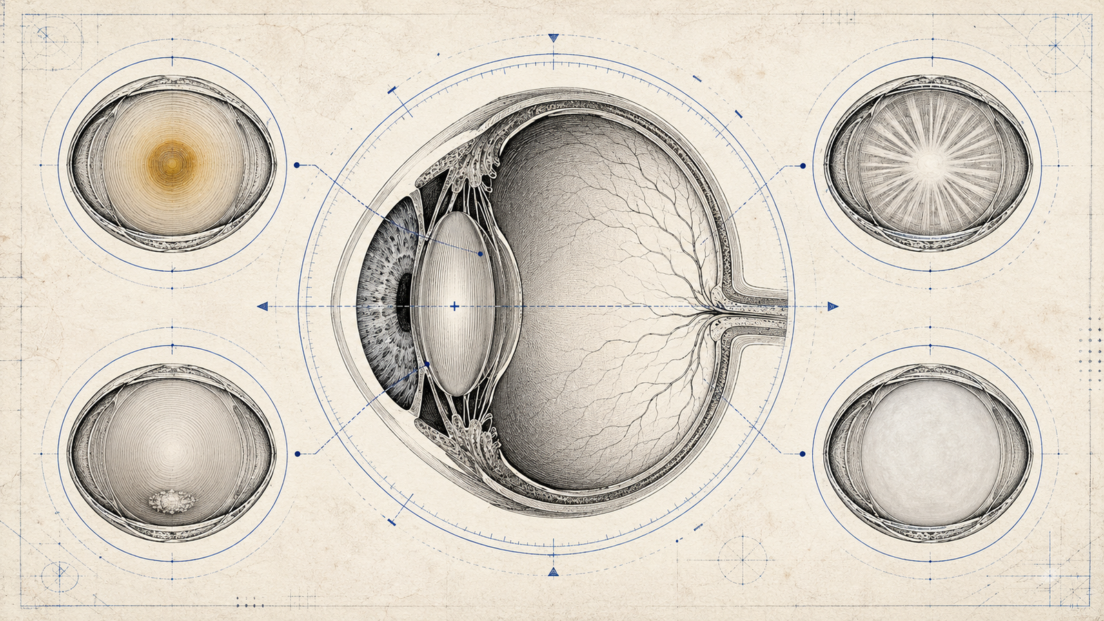
| Nuclear sclerosis | Progressive yellowing/hardening of the lens nucleus; classically causes a myopic shift, sometimes described by patients as temporarily improved near vision — the "second sight" phenomenon |
| Cortical | Spoke-like or wedge-shaped opacities radiating from the periphery toward the center |
| Posterior subcapsular | Opacity just in front of the posterior lens capsule; disproportionately symptomatic for its size |
Why Posterior Subcapsular Cataracts Punch Above Their Size
Because they sit directly at the visual axis — right where light converges — even a small posterior subcapsular opacity causes disproportionate symptoms: glare, particularly in bright light, and difficulty with near vision and reading. This is also the type most associated with steroid use and diabetes, which is a frequently tested pairing. The visual axis location, rather than the absolute size of the opacity, is what determines symptom severity here — a detail worth internalizing, since it's a genuinely counterintuitive point that a question can exploit.
The "Second Sight" Phenomenon, Explained
As the lens nucleus hardens and its refractive index increases in nuclear sclerosis, the eye's overall refractive power increases — inducing a myopic shift. Some older patients notice they can temporarily read without their reading glasses as this happens, before the cataract progresses further and vision worsens overall. It's a genuinely useful clinical clue, not just a curiosity, since a patient reporting sudden, unexplained improvement in near vision without new correction is a classic setup for this exact phenomenon.
Congenital Cataract: A Distinct, High-Stakes Category
Congenital cataract deserves separate attention because early recognition has real functional consequences — an untreated congenital cataract can cause irreversible amblyopia if not addressed within the critical period of visual development in infancy. The classic causes worth knowing are intrauterine infections, particularly rubella (part of the TORCH group), along with galactosemia and various inherited metabolic disorders. A white pupillary reflex (leukocoria) noticed in a newborn or young infant is a red flag finding that should prompt urgent ophthalmologic evaluation, since congenital cataract is one of several serious causes of leukocoria alongside retinoblastoma — a distinction the exam expects you to hold in mind rather than assume leukocoria always means one specific diagnosis.
Complications Worth Knowing
An untreated, advanced (hypermature) cataract can lead to two specific complications worth knowing by name. Phacolytic glaucoma occurs when lens proteins leak through an intact but permeable capsule, triggering an inflammatory response that obstructs the trabecular meshwork and raises intraocular pressure. Lens-induced (phacoanaphylactic) uveitis occurs when the immune system reacts against exposed lens proteins, typically after capsular rupture from trauma or surgery. Both are reminders that a cataract isn't always just a passive visual disturbance — an advanced or ruptured lens can become an active source of ocular inflammation and pressure elevation in its own right.
Cataract Surgery: The Basic Concept Worth Knowing
Modern cataract surgery is most commonly performed via phacoemulsification — using ultrasound energy to break up and remove the cloudy lens through a small incision — followed by implantation of an intraocular lens (IOL) to restore refractive power. Surgery is generally indicated once the cataract significantly impairs a patient's functional vision or quality of life, rather than at a fixed severity threshold, since the decision is ultimately guided by the patient's actual visual needs rather than a specific test result alone.
A Few Pairings Worth Double-Checking
- Steroid use and posterior subcapsular cataract is one of the most frequently tested cause-to-morphology pairings in this topic.
- Diabetic ("snowflake") cataract develops through a distinct osmotic mechanism (sorbitol accumulation in the lens), separate from the mechanism behind ordinary age-related cataract, even though diabetic patients also develop age-related cataracts earlier and more commonly than non-diabetics.
- Rubella and congenital cataract is a classic association worth remembering alongside the other features of congenital rubella syndrome (which also include congenital heart defects and sensorineural hearing loss) — a triad that shows up together often enough to be worth learning as a set.
Cataract Maturity: The Staging Terminology
Beyond cause and morphology, cataracts are also staged by maturity, and this terminology carries real clinical implications worth knowing precisely. An immature cataract shows partial lens opacity with some remaining clear cortex, and the red reflex is typically still partially visible on examination. A mature cataract shows complete opacification of the lens, with the red reflex absent. A hypermature cataract represents a further stage where the lens has begun to liquefy and shrink, with a wrinkled anterior capsule — and this is the stage most associated with the complications discussed above, since liquefied lens material is what leaks through the capsule to cause phacolytic glaucoma. A Morgagnian cataract is an advanced hypermature form where the liquefied cortex allows the dense nucleus to sink within the lens capsule, visible as a distinctive downward displacement on examination. Recognizing where a described cataract sits on this maturity spectrum often clarifies both the expected exam findings and the urgency of intervention.
Unilateral vs Bilateral: A Clue Worth Using
Whether a cataract is unilateral or bilateral is itself diagnostically useful, not just an incidental detail. Bilateral cataracts are more typical of systemic causes — age-related change, metabolic conditions like diabetes or galactosemia, and congenital causes like intrauterine rubella infection, since these all affect both eyes through a shared systemic mechanism. A unilateral cataract more often points toward a localized cause specific to that eye — trauma, or a complicated cataract secondary to chronic uveitis or high myopia confined to one eye. When a vignette specifies laterality, it's often doing so deliberately, nudging you toward a systemic versus localized cause rather than leaving you to guess from the morphology alone.
Cause-to-appearance matching like this is exactly the pattern Reflex's daily TROCAR Ophthalmology rounds are built to reinforce.
Practise this on Reflex
Turn what you just read into recall with 14 years of tagged PYQs.
FAQ
Frequently asked questions
The questions aspirants ask most about this topic.
Age-related changes are the most common cause overall; other causes include congenital factors, trauma, metabolic conditions (diabetes, galactosemia), and drugs such as corticosteroids.
A temporary improvement in near vision caused by the myopic shift from a hardening, more refractive lens nucleus in early nuclear sclerosis — before the cataract progresses further and vision worsens overall.
Posterior subcapsular cataract.
Because they're located directly at the visual axis, where light converges, so even a small opacity there disproportionately affects vision — especially glare and near vision.
A white pupillary reflex, which is a red-flag finding in infants requiring urgent evaluation — congenital cataract and retinoblastoma are both serious causes that must be distinguished promptly.
A complication of an advanced (hypermature) cataract, where leaking lens proteins trigger inflammation that obstructs the trabecular meshwork, raising intraocular pressure.
An advanced hypermature cataract where the liquefied cortex allows the dense lens nucleus to sink within the capsule, visible as a distinctive downward displacement on examination.
An immature cataract has partial lens opacity with some clear cortex remaining and a partially visible red reflex. A mature cataract shows complete opacification with an absent red reflex — a distinction that also signals how far the cataract has progressed toward the complications seen in hypermature disease.
Often, yes. Bilateral cataracts more typically reflect a systemic cause (age-related change, diabetes, galactosemia, congenital rubella), while a unilateral cataract more often points toward a localized cause specific to that eye, such as trauma or chronic uveitis confined to one side — a distinction worth actively considering whenever a question specifies which pattern is present.
About the author
Stay ahead in your preparation
Get expert tips, exam updates, and high-yield insights delivered straight to your inbox.
No spam. Unsubscribe anytime.
More in Subject Notes
View all → Ophthalmology