Types of Fractures for NEET PG: The Two Axes Every Question Actually Tests
By Dr. Utsav Bhattacherjee, MBBS, MBA · 21 Sept 2026 · 13 min read
Last updated: 23 Sept 2026
On this page
Fracture classification questions are built around two separate axes that get tested together constantly: the pattern of the break itself, and whether the skin over it is intact. Keep those two axes separate in your head, and most fracture questions become a matter of matching the description to the right box rather than genuine recall difficulty.
Classification by Pattern
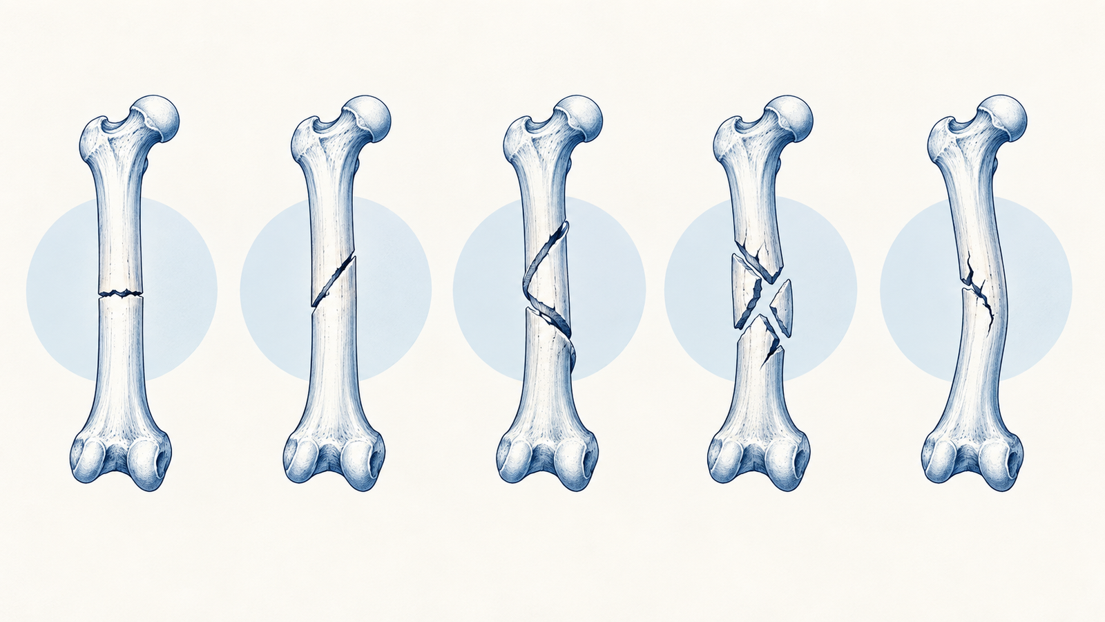
A transverse fracture runs straight across the bone, perpendicular to its long axis, and typically results from a direct blow. An oblique fracture runs at an angle across the bone, usually from an angulated force. A spiral fracture wraps around the bone in a helical pattern and is the classic signature of a rotational or twisting force — its presence in a young child, without a matching mechanism in the history, is a well-known red flag for non-accidental injury.
A comminuted fracture shatters the bone into three or more fragments, typically from high-energy trauma, and carries a worse healing prognosis simply because there are more fragment interfaces that need to unite. A greenstick fracture is an incomplete break where one side of the bone bends and cracks while the other side stays intact — a pattern specific to the more pliable, incompletely mineralized bone of children, and essentially never seen in adult bone. A compression fracture results from an axial loading force crushing the bone, classically seen in vertebral bodies in osteoporotic patients.
Open vs Closed: Why This Axis Is Separate From Pattern
A closed (simple) fracture has intact overlying skin. An open (compound) fracture has a break in the skin communicating with the fracture site, and this distinction matters enormously beyond just description — an open fracture is a surgical emergency carrying real infection risk, regardless of how simple or comminuted the underlying bone pattern is. A transverse fracture and a comminuted fracture can each be open or closed independently; the two classification systems describe genuinely different things about the same injury.
The Gustilo-Anderson Classification for Open Fractures
Open fractures are further graded by the Gustilo-Anderson system, which correlates with infection risk and guides management urgency. Type I involves a wound under 1cm with minimal soft tissue damage. Type II involves a wound over 1cm with moderate soft tissue damage but no extensive flap or avulsion. Type III is the most severe category, further split into IIIA (adequate soft tissue coverage despite extensive damage), IIIB (extensive soft tissue loss requiring flap coverage), and IIIC (an open fracture with an associated arterial injury requiring repair) — IIIC carries the highest infection and amputation risk of the entire system.
Named Fractures Worth Knowing by Sight
Certain fractures carry eponymous names specifically because their mechanism, pattern, and management are distinct enough to warrant separate recognition. A Colles' fracture is a distal radius fracture with dorsal (backward) displacement of the fragment, classically resulting from a fall onto an outstretched hand, producing the characteristic dinner-fork deformity on examination. A Smith's fracture is essentially its mirror image — a distal radius fracture with volar (forward) displacement, typically from a fall onto a flexed wrist.
A Monteggia fracture combines a fracture of the proximal ulna with dislocation of the radial head — the two injuries occur together because the interosseous membrane and annular ligament connecting the radius and ulna transmit force between them. A Galeazzi fracture is the mirror pairing: a fracture of the distal radius combined with dislocation of the distal radioulnar joint. Mixing up which bone fractures in each (ulna in Monteggia, radius in Galeazzi) is a common, specifically testable error worth guarding against directly.
A Pott's fracture describes a bimalleolar or trimalleolar ankle fracture, classically from an external rotation and eversion force at the ankle. Recognizing these named patterns quickly, rather than reconstructing the anatomy from scratch each time, is exactly what separates fast, confident answers from ones reasoned out under time pressure.
| Named Fracture | Bone(s) Involved | Classic Mechanism |
|---|---|---|
| Colles | Distal radius, dorsal displacement | Fall on outstretched hand |
| Smith | Distal radius, volar displacement | Fall on flexed wrist |
| Monteggia | Proximal ulna + radial head dislocation | Direct blow or fall with forearm rotation |
| Galeazzi | Distal radius + distal radioulnar joint dislocation | Fall on outstretched hand with forearm rotation |
| Pott | Bimalleolar / trimalleolar ankle | External rotation and eversion |
Stress Fractures and Pathological Fractures: Two Different Reasons Bone Fails
A stress fracture results from repetitive, cumulative loading on normal bone rather than a single traumatic event — classically seen in the metatarsals of runners or military recruits undergoing sudden increases in training load. The bone itself is structurally normal; the failure comes from repeated microtrauma outpacing the bone's normal remodeling capacity.
A pathological fracture, by contrast, occurs in bone that is already structurally weakened by an underlying condition — a bone tumour, a metastatic deposit, osteoporosis, or a metabolic bone disease — breaking under a force that would not fracture normal bone. A vignette describing a fracture from a trivial mechanism (a minor fall, or even normal daily activity) should immediately raise the question of what underlying process weakened the bone in the first place, rather than treating it as an unusually severe injury from a minor cause.
Fracture Healing and Why Certain Fractures Heal Poorly
Fracture healing proceeds through a hematoma formation stage, a soft callus stage as fibrocartilage bridges the fracture gap, a hard callus stage as this is mineralized into woven bone, and finally remodeling into mature lamellar bone over months. Certain fractures are well known to heal poorly or slowly specifically because of compromised blood supply to one fragment — the scaphoid (particularly proximal pole fractures), the femoral neck, and the talus are the classic examples, each vulnerable because their blood supply enters at one end and can be disrupted by the fracture itself, leaving the other fragment without adequate perfusion to heal normally.
Fracture Descriptions on Imaging: The Language Worth Knowing
Beyond naming the pattern itself, fracture descriptions on imaging follow a standard vocabulary worth recognizing. Displacement describes the fragments shifting sideways relative to each other, described as a percentage of bone width. Angulation describes the fragments tilting relative to each other, described in degrees and by the direction the distal fragment points. Shortening describes the fragments overlapping along the bone's long axis, reducing overall bone length. Rotation describes one fragment twisting relative to the other along the bone's long axis, which can be surprisingly easy to miss on a single X-ray view and is exactly why fracture imaging is taken in at least two perpendicular planes rather than one.
This is also why a fracture that looks straightforward on one view can reveal significant rotational deformity on the perpendicular view, changing management from conservative casting to a discussion about surgical fixation — the single-view description in a vignette is often deliberately incomplete, testing whether the reader knows to ask what the second view would show rather than accepting the first image as the whole picture.
Reading a fracture vignette efficiently means checking pattern, skin status, and mechanism in that order: the pattern narrows toward a named fracture or general category, skin status determines whether this is a surgical emergency regardless of pattern, and mechanism either confirms a trivial-force pathological fracture is hiding underneath, or points toward a specific named injury the description is building toward.
For the same pattern-recognition approach applied to a related orthopedic topic, see our guide to Salter-Harris fractures, and for how much of the paper Orthopedics carries overall, our subject-wise weightage breakdown.
Practise this on Reflex
Turn what you just read into recall with 14 years of tagged PYQs.
FAQ
Frequently asked questions
The questions aspirants ask most about this topic.
A comminuted fracture shatters into three or more fragments at one site. A segmental fracture has two separate fracture lines creating a distinct, free-floating middle segment of bone.
Spiral fractures result from a rotational or twisting force. In a young child without a matching mechanism in the history, it raises concern for non-accidental injury.
It involves an associated arterial injury requiring surgical repair, on top of the soft tissue damage seen in other Type III fractures.
A Monteggia fracture involves the proximal ulna with radial head dislocation. A Galeazzi fracture involves the distal radius with distal radioulnar joint dislocation.
Both have blood supply entering at one end of the bone, which the fracture itself can disrupt, leaving the other fragment without adequate perfusion to heal normally.
A stress fracture occurs in structurally normal bone from repetitive loading. A pathological fracture occurs in bone already weakened by an underlying condition, breaking under a force that would not fracture normal bone.
About the author
Stay ahead in your preparation
Get expert tips, exam updates, and high-yield insights delivered straight to your inbox.
No spam. Unsubscribe anytime.
More in Subject Notes
View all → Orthopedics
Salter-Harris Classification for NEET PG: The SALTR Mnemonic, Explained Properly
Salter-Harris questions test two things: reading the type off an X-ray description, and knowing which type carries the worst prognosis. The SALTR mnemonic, why Type V is the trap, and how management differs across the five types.
Malaria Life Cycle for NEET PG: Species, Smear Morphology, Complications and Treatment
How the malaria parasite moves between mosquito and human, what separates the species on a smear, and how diagnosis, complications and treatment follow from the life cycle.
Oxygen-Hemoglobin Dissociation Curve for NEET PG: Every Shift Explained
Why the oxygen-hemoglobin dissociation curve is sigmoid, what moves it right or left, and how to apply each shift to fetal hemoglobin, stored blood, altitude and carbon monoxide poisoning.
Cholinergic Drugs for NEET PG: Classification, Receptors and Organophosphate Poisoning
A working classification of cholinergic drugs, the receptors behind each effect, which drug fits which clinical scenario, and how to answer organophosphate poisoning questions.