Salter-Harris Classification for NEET PG: The SALTR Mnemonic, Explained Properly
By Dr. Utsav Bhattacherjee, MBBS, MBA · 27 Aug 2026 · 10 min read
Last updated: 23 Sept 2026
On this page
Salter-Harris fracture questions in Orthopedics almost always test one of two things: identifying the type from an X-ray description, or knowing which type carries the worst prognosis — and the two aren't always the answer you'd instinctively expect.
Why Growth Plate Fractures Are Their Own Category
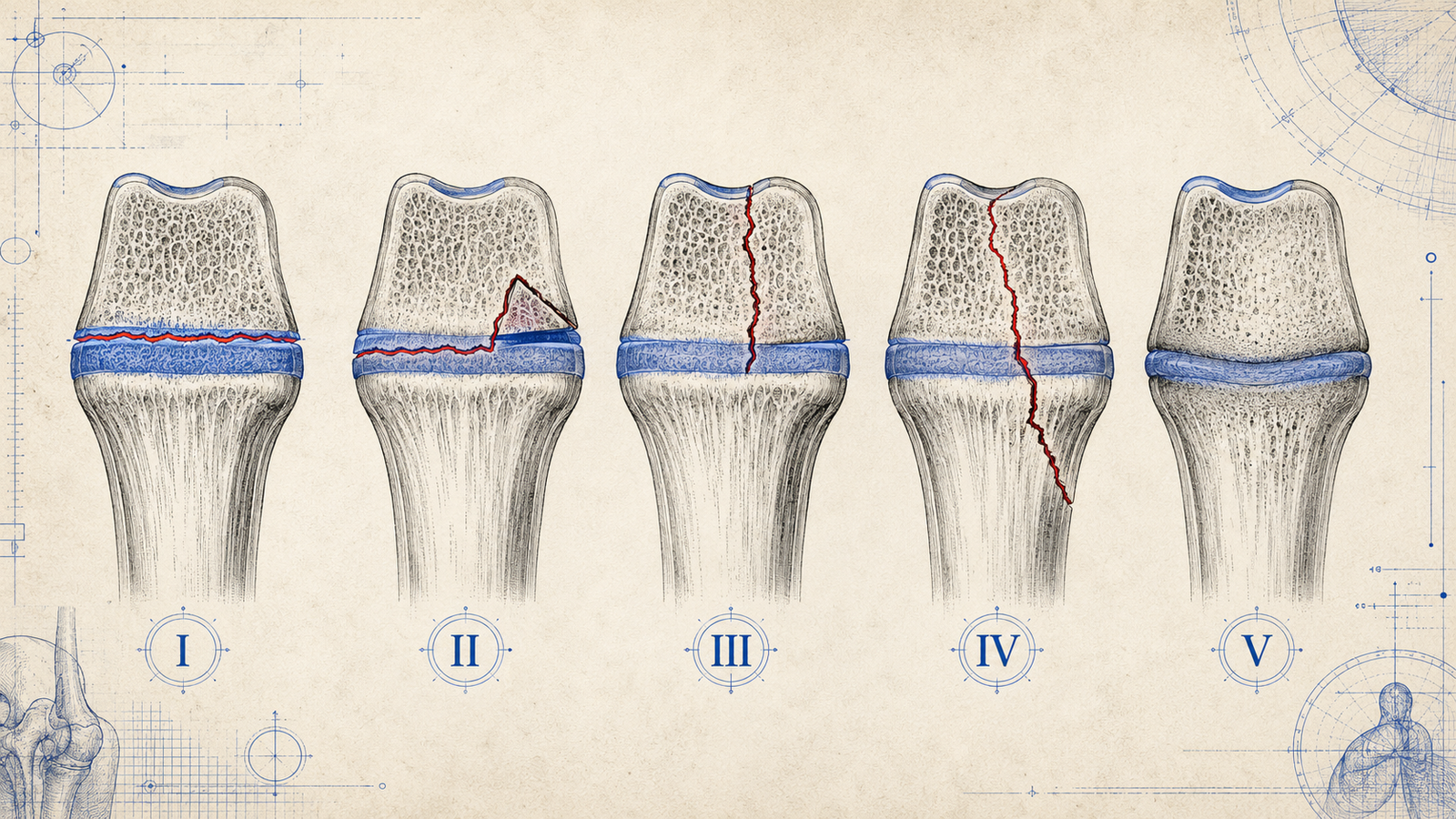
Children's bones have a feature adults' bones don't: the physis, or growth plate, a layer of cartilage near each end of a long bone where growth occurs. This makes pediatric fractures involving the growth plate a genuinely distinct category from adult fracture patterns, since damage here carries a risk adult fractures simply don't — the potential for growth arrest or angular deformity as the child continues to grow. The Salter-Harris system exists specifically to classify these growth-plate-involving fractures by where the fracture line runs relative to the physis, because that location is what predicts the risk to future growth.
The SALTR Mnemonic
| Type | Letter | Meaning | Fracture line |
|---|---|---|---|
| I | S | Slipped | Along the physis (growth plate) only |
| II | A | Above | Extends above the physis, into the metaphysis |
| III | L | Lower (beLow) | Extends below the physis, into the epiphysis |
| IV | T | Through | Extends through metaphysis, physis, and epiphysis |
| V | R | Rammed (cRush) | Crush injury to the growth plate itself |
Which Type Is Most Common
Type II is the most common Salter-Harris fracture overall, accounting for the majority of growth plate injuries. Its fracture line extends above the physis into the metaphysis, sparing the epiphysis and joint surface — a pattern that generally carries a favorable prognosis with proper management.
Which Type Has the Worst Prognosis
This is the classic exam twist: Type V, the crush injury, carries the worst prognosis for growth disturbance — despite often looking unremarkable, or even normal, on initial X-ray. The growth plate can be significantly damaged by the crushing mechanism without an obvious fracture line, which makes it easy to under-diagnose acutely and only recognize later, once growth arrest becomes apparent. The exam-relevant lesson: a normal-looking initial X-ray in the right clinical context (a crush mechanism of injury) shouldn't be taken as reassurance that the growth plate is undamaged.
Why This Classification Matters Clinically, Not Just for the Exam
Growth plate injuries carry a risk the same fracture wouldn't carry in a skeletally mature adult. Types I and II generally carry a good prognosis with proper reduction, since they don't involve the epiphysis or joint surface directly. Types III and IV, involving the epiphysis, carry higher risk of both growth disturbance and joint surface incongruity, since the fracture disrupts the articular surface as well as the growth mechanism. Type V carries the highest overall risk of growth arrest, precisely because the crushing mechanism damages the growth plate's cellular architecture directly, regardless of how the bone ends look on imaging.
Management Principles by Type
Types I and II are generally managed with closed reduction and casting, since the fracture pattern doesn't typically disrupt the joint surface and the growth plate's own healing capacity is often sufficient once properly aligned. Types III and IV, given their involvement of the joint surface, more frequently require open reduction and internal fixation to precisely restore both the joint surface and physeal alignment — anatomic reduction matters more here because any residual displacement risks both joint incongruity and asymmetric growth. Type V has no truly effective definitive treatment for the crush injury itself, since the damage is often done at the moment of injury — management focuses on monitoring for growth arrest and managing any resulting deformity if it develops, which is part of why this type's prognosis is considered the worst of the five regardless of how promptly it's recognized.
Comparing Salter-Harris to Adult Fracture Description
It's worth explicitly contrasting this system with how fractures are typically described in adults, since conflating the two frameworks is a common source of confusion. Adult fracture description focuses on pattern (transverse, oblique, spiral, comminuted), displacement, and angulation — descriptive terms that apply regardless of age. Salter-Harris classification is specifically about the fracture's relationship to the growth plate, a structure that doesn't exist in the same way once growth plates fuse in early adulthood. A pediatric fracture can be described using both frameworks simultaneously (a Type II fracture that is also, say, oblique and minimally displaced) — the two systems aren't competing, they're answering different questions about the same injury.
A Common Exam Trap Worth Naming
Because Type II is both the most common and generally has a good prognosis, it's tempting to assume "common" and "concerning" don't overlap in this topic. Type V demonstrates exactly why that assumption is dangerous: a genuinely rare fracture type, easily missed on initial imaging, carries the single worst long-term outcome of the five. A question that describes a crush mechanism of injury with an unremarkable X-ray is very likely testing whether you'll correctly suspect Type V despite the reassuring-looking image, rather than assuming a normal film rules out significant growth plate injury.
Other Pediatric-Specific Fracture Patterns Worth Knowing
Salter-Harris isn't the only fracture pattern unique to children — a couple of others are worth knowing alongside it, since pediatric bone is more pliable than adult bone and fails differently under stress. A greenstick fracture is an incomplete fracture where the bone bends and cracks on one side (the tension side) while remaining intact on the other (the compression side) — named for its resemblance to bending a green, still-flexible twig rather than a dry one that snaps cleanly. A buckle (torus) fracture is a compression injury where the bone buckles outward without a clear fracture line crossing the cortex, typically from a fall onto an outstretched hand, and is generally a stable, low-risk injury. Neither of these involves the growth plate directly, which is exactly why they're classified separately from the Salter-Harris system — they're a reminder that "pediatric fracture" is a broader category than growth-plate injuries alone, even though Salter-Harris gets the most exam attention given its direct implications for future growth. Both greenstick and buckle fractures also illustrate the same underlying principle that makes pediatric bone behave differently from adult bone in the first place: a child's skeleton is more porous and has a thicker, more active periosteum, giving it greater flexibility before it fails — which is exactly why children bend and buckle where an adult of comparable force would more often sustain a complete fracture.
Classification-and-prognosis questions like this are exactly what Reflex's daily TROCAR Orthopedics rounds are built to reinforce.
Practise this on Reflex
Turn what you just read into recall with 14 years of tagged PYQs.
FAQ
Frequently asked questions
The questions aspirants ask most about this topic.
Slipped (Type I), Above (Type II), Lower/beLow (Type III), Through (Type IV), and Rammed/cRush (Type V) — describing where the fracture line runs relative to the growth plate.
Type II.
Type V, the crush injury, despite frequently appearing unremarkable on initial imaging.
Because damage to the growth plate can cause growth arrest or angular deformity as the child continues to grow — a risk that doesn't apply to the same fracture pattern in a skeletally mature adult.
Often yes — since they involve the epiphysis and joint surface, open reduction and internal fixation is more frequently required to restore precise joint and physeal alignment, compared to Types I and II, which are more often managed with closed reduction.
Yes — this is exactly what makes it dangerous. The crush mechanism can significantly damage the growth plate without producing an obvious fracture line, so a normal-looking initial film doesn't rule out significant injury in the right clinical context.
An incomplete pediatric fracture where the bone bends and cracks on the tension side while remaining intact on the compression side — distinct from Salter-Harris fractures since it doesn't involve the growth plate.
No. A buckle fracture is a compression injury where the bone buckles without a clear fracture line, typically from a fall onto an outstretched hand, and doesn't involve the growth plate — it's a separate, generally stable pediatric fracture pattern classified outside the Salter-Harris system.
About the author
Stay ahead in your preparation
Get expert tips, exam updates, and high-yield insights delivered straight to your inbox.
No spam. Unsubscribe anytime.
More in Subject Notes
View all → Orthopedics