Visual Pathway and Its Lesions for NEET PG: A Site-by-Site Field Defect Map
Reflex · 2 Nov 2026 · 12 min read
Last updated: 2 Nov 2026
On this page
Every visual field question is a localisation question. The defect on the perimetry chart tells you where along the visual pathway the damage sits, and the pathway is short enough to learn as a single map.
This post walks from retina to occipital cortex, asks one question at each station (what has been cut, and what does the patient lose?), and ends with a table you can redraw from memory. It also covers the points where textbooks disagree, such as how reliable the congruity rule is and why the macula is spared in some occipital lesions.
If you are working through the rest of Ophthalmology, the red eye comparison pairs well with this topic.
Key Takeaways
Nasal retinal fibres (which see the temporal field) cross at the chiasm; temporal retinal fibres stay on the same side. Each optic tract therefore carries the opposite half of the visual world.
Before the chiasm, a lesion causes loss in one eye. At the chiasm, think bitemporal hemianopia. After the chiasm, think homonymous defects on the opposite side.
Temporal lobe (Meyer loop) lesions give a superior quadrantanopia; parietal lesions give an inferior quadrantanopia.
The farther back the lesion, the more congruous the defect tends to be, but this is a tendency and not a rule.
Occipital lesions classically give a congruous homonymous hemianopia, often with macular sparing; the reason for sparing is still debated.
A tract lesion can produce a relative afferent pupillary defect in the eye with the temporal field loss. A marked one is not expected with post-geniculate lesions.
How the Visual Pathway Runs From Retina to Cortex
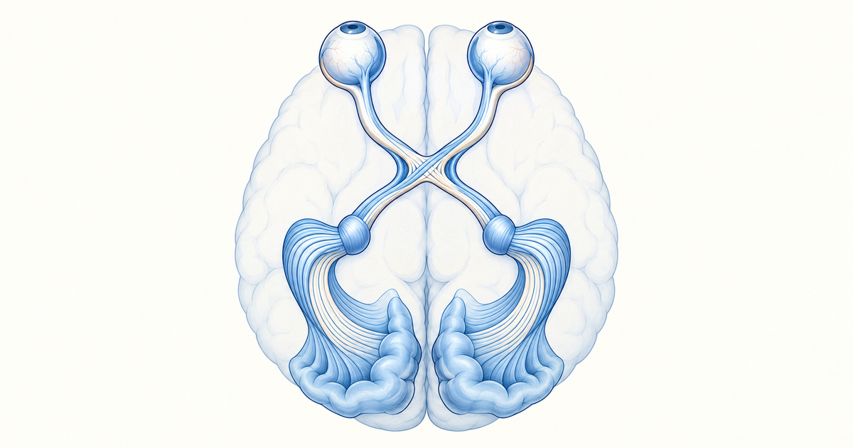
The visual pathway is a relay of five stations: retina, optic nerve and chiasm, optic tract, lateral geniculate nucleus (LGN), and optic radiation to the occipital cortex. Patel and colleagues describe this course: photoreceptors pass signals to bipolar cells, bipolar cells to ganglion cells, and ganglion cell axons form the optic nerve. The two nerves meet at the chiasm, the tracts run on to the LGN, and the radiations travel through the internal capsule to the visual cortex.
Which fibres cross at the chiasm
Nasal retinal fibres cross at the chiasm and temporal retinal fibres do not. The nasal fibres carry the temporal visual field, which is why the crossing matters. The same source adds that each optic tract therefore carries the opposite hemifield: the right tract carries the left half of the visual world from both eyes.
A quick drill: the left tract carries the right visual field, drawn from the left temporal retina (uncrossed) and the right nasal retina (crossed). If you can say that without hesitation, most lesion questions become mechanical.
Which radiation carries which part of the field
The optic radiation splits by the part of the field it serves. According to the same review, superior-field fibres loop through the temporal lobe (Meyer loop) to the lower lip of the calcarine sulcus, while inferior-field fibres run through the parietal lobe to the upper lip.
Radiopaedia's radiation article describes the same anatomy slightly differently: the radiation passes through the retrolentiform part of the internal capsule and fans into bundles. The anterior bundle (Meyer loop) curves around the roof of the temporal horn, and the posterior bundle runs back to the superior lip of the calcarine sulcus. Some books call the posterior bundle the parietal radiation, so treat the two names as the same structure.
A memory aid built from these facts (not a published mnemonic): temporal lobe, top of the field; parietal lobe, bottom of the field.
Lesion-to-Field-Defect Map Along the Visual Pathway
The table below is the centrepiece of this post. Learn it as one picture, from front to back, and notice that the defect changes character at two boundaries: the chiasm and the LGN.
Sources for this table: Patel et al., Radiopaedia on visual pathway deficits, and EyeWiki on compressive field defects.
| Site of lesion | Field defect | Key point |
|---|---|---|
| Optic nerve | Loss of vision in the same eye, or a central scotoma | Monocular |
| Chiasm, middle (classic) | Bitemporal hemianopia | Crossing nasal fibres compressed; often asymmetric |
| Chiasm, anterior | Junctional scotoma: central scotoma in the ipsilateral eye plus a superotemporal defect in the other eye | Anterior chiasm lesion; the "one-eyed" clue misleads |
| Chiasm, posterior | Scotomatous bitemporal hemianopia | Posterior chiasm lesion |
| Chiasm, lateral | Binasal or ipsilateral nasal defect | Described as uncommon |
| Optic tract | Contralateral homonymous hemianopia | Classically incongruous |
| LGN | Homonymous sectoranopia or hemianopia | Rare lesion site |
| Temporal radiation (Meyer loop) | Contralateral homonymous superior quadrantanopia | "Pie in the sky" |
| Parietal radiation | Contralateral homonymous inferior quadrantanopia | "Pie on the floor" |
| Occipital cortex | Contralateral congruous homonymous hemianopia | Macula often spared |
| Both occipital lobes | Cortical blindness | Bilateral lesion |
Two rules sit behind the whole table. First, a lesion in front of the chiasm affects one eye. Second, a lesion behind the chiasm affects the opposite half of the field in both eyes, which is what "homonymous" means.
Optic Chiasm Lesions and Bitemporal Hemianopia
Bitemporal hemianopia is the signature of a chiasmal lesion. The MSD Manual lists it for chiasmal disease and names the usual culprits: pituitary tumour, craniopharyngioma, meningioma, glioma and aneurysm. EyeWiki adds that pituitary adenoma is the most common neoplastic cause.
The mechanism follows from the anatomy. A mass pressing on the middle of the chiasm squeezes the decussating nasal fibres, which carry the temporal fields, so both temporal fields go. The defect is often asymmetric, as noted in the Patel review above.
Anterior and posterior chiasm patterns
Not every chiasmal lesion gives a clean bitemporal hemianopia. Radiopaedia separates anterior, middle and posterior chiasmal lesions. An anterior lesion produces a junctional scotoma, a posterior one gives a scotomatous bitemporal hemianopia, and the middle one gives the classic form.
EyeWiki describes the junctional scotoma of Traquair as a defect in one eye with an ipsilateral temporal defect caused by compression of crossing nasal fibres, with the other eye spared. Use that as a reminder that "one-eyed" field loss does not always mean the optic nerve alone.
Binasal defects
Binasal hemianopia is uncommon. The Patel review and Radiopaedia link it with lateral or asymmetric chiasmal lesions, and the MSD Manual table lists it as uncommon. Do not guess binasal when you see a bitemporal picture, because the two are mirror images.
Visual Pathway Lesions Behind the Chiasm
Behind the chiasm, the defect becomes homonymous: the same half of the field is lost in both eyes. MSD notes that a homonymous hemianopia can arise from a lesion anywhere behind the chiasm, usually stroke or tumour. In adults, StatPearls reports cerebral infarct and intracranial haemorrhage as the commonest causes, with tumour and trauma next.
The same StatPearls chapter gives a lesion-site breakdown from one large series: occipital lobe 45%, optic radiation 32.2%, optic tract 10.2% and LGN 1.3%. Treat these as figures from a single series and not as universal rates.
Optic tract and LGN
A tract lesion gives a contralateral homonymous hemianopia, classically incongruous. LGN lesions are rare, and Patel et al. list a homonymous sectoranopia or hemianopia, with ischaemic, demyelinating or traumatic causes.
Temporal and parietal radiations
Here the quadrant tells you the lobe. A superior quadrantanopia ("pie in the sky") points to the temporal lobe because Meyer loop carries the superior field. An inferior quadrantanopia ("pie on the floor") points to the parietal lobe.
EyeWiki lists extra clues that help localise the lobe. Temporal lesions may bring aphasia or hallucinations, a left parietal lesion may cause Gerstmann syndrome, and a right parietal lesion may cause neglect. Bilateral parietal damage may cause Balint syndrome.
Self-test: name the lesion
A patient cannot see the upper right quadrant of the visual field in either eye. Which lobe is involved? The left temporal lobe, because the right visual field is carried by the left hemisphere and the superior field passes through Meyer loop.
Occipital Lesions and Macular Sparing in the Visual Pathway
An occipital lesion classically produces a congruous homonymous hemianopia, usually with macular sparing. EyeWiki and Patel et al. both describe this, with a posterior cerebral artery (PCA) stroke as the textbook example. Macular sparing means a small central area of vision survives inside the blind hemifield.
Why the macula is spared
The commonly taught explanation is vascular: the occipital pole has a dual supply from the PCA and middle cerebral artery (MCA), so a PCA occlusion may leave the macular representation perfused. StatPearls lists four theories in all. The others are a large cortical area devoted to the central field, anastomotic blood supply at the occipital poles, and representation of the macula in both occipital lobes.
Present the mechanism as debated. What is not in doubt is the pattern: preserved central field within a dense hemianopia points posteriorly. The same chapter notes that macular sparing can also occur with radiation or tract lesions, so it is a pointer and not proof.
Bilateral occipital lesions
When both occipital lobes are damaged, the result is cortical blindness, as the Patel review notes. Radiopaedia also lists Anton and Balint syndromes for bilateral occipital and occipitoparietal lesions.
Congruity: A Tendency, Not a Rule
Congruity describes how closely the defects in the two eyes match in shape and size. The familiar teaching is that the farther back the lesion, the more congruous the defect. Tract lesions are classically incongruous, and occipital lesions are congruous.
The real data are messier. StatPearls reports that about half of tract lesions are congruous, around 60% of radiation lesions are congruous, and about 17% of occipital lesions are still incongruous. Kedar and colleagues report that almost half of tract lesions give a congruent hemianopia, and that about 56% of congruent hemianopias come from occipital lesions.
So use congruity as supporting evidence. A markedly incongruous hemianopia leans toward the tract or anterior radiation, a perfectly congruous one leans occipital, and neither is a guarantee.
Pupil Signs and the Visual Pathway
Pupil fibres leave the visual pathway at the optic tract and exit at the brachium of the superior colliculus, so the pupil helps distinguish a tract lesion from lesions further back. Bell and Thompson described a relative afferent pupillary defect (RAPD) in the eye with the temporal field loss in optic tract hemianopias, which is the eye opposite the lesion. They found it in all four patients with a complete tract hemianopia.
EyeWiki adds that band atrophy of the optic disc may appear in the eye with the temporal defect.
What happens with post-geniculate lesions
A clinically obvious RAPD points to the anterior pathway or the tract. Post-geniculate lesions typically do not produce a marked RAPD, though sensitive pupillometry can detect a small one. Takizawa and colleagues found a small pupillometry-detected RAPD (about 0.13 log units) in 15 patients with occipital stroke, compared with about 0.49 log units in tract syndrome.
The light reflex itself runs along a separate route, as this review outlines: the retinomesencephalic tract carries signals to the midbrain pretectal area, then to both Edinger-Westphal nuclei and cranial nerve III.
Testing the Visual Pathway: Confrontation to Perimetry
Confrontation testing is the bedside screen, but it is not sensitive enough to rule out a defect. StatPearls describes it as an insensitive method whose sensitivity may improve with a red target. In the review by Kedar et al., confrontation detected only about 20% of arcuate defects and about 70% of hemianopias, and roughly 75% with a small red target. When a positive result is confirmed by automated perimetry, specificity is high (97%).
Static automated perimetry (Humphrey 10-2, 24-2 or 30-2) is recommended for unexplained visual loss, according to EyeWiki, and has largely replaced Goldmann perimetry in neuro-ophthalmology. The Kedar review adds that static perimetry can show temporal depression and a vertical step early in chiasmal lesions, while Goldmann kinetic testing is kept for severe deficits and isolated peripheral defects. To test for right parietal neglect, EyeWiki suggests simultaneous confrontation of both fields.
Once the theory is clear, practise field-chart localisation with the neuro-ophthalmology practice questions.
Common Mistakes When Localising Visual Pathway Lesions
These errors come from mixing up the rules, and each has a quick fix.
- Calling a monocular defect "chiasmal". A defect confined to one eye usually means a lesion in front of the chiasm, except for the junctional scotoma, which also has a contralateral superotemporal component.
- Swapping the quadrants. Temporal lobe means superior field loss; parietal lobe means inferior field loss.
- Treating congruity as proof. An incongruous defect does not exclude an occipital lesion, as the series above show.
- Reading macular sparing as a diagnosis. It points posteriorly but can also occur with tract or radiation lesions.
- Expecting a pupil defect in every hemianopia. Look for a clinically obvious RAPD with tract lesions, not with typical post-geniculate lesions.
Self-test: bitemporal loss
A patient has bitemporal hemianopia. Which fibres are being compressed? The crossing nasal retinal fibres at the middle of the chiasm, so the first thought is a sellar or suprasellar mass such as a pituitary adenoma. Test yourself again by covering the table above and redrawing it from memory.
Conclusion
The visual pathway is easiest to learn as a sequence of decisions: one eye or both, crossing or non-crossing fibres, which quadrant, and whether the macula survives. Hold on to the chiasm and the LGN as the two boundaries where the pattern changes, and use congruity and pupil signs as supporting clues rather than proof.
Redraw the lesion table from memory until it is automatic, then move on to another Ophthalmology topic such as cataract types.
Practise this on Reflex
Turn what you just read into recall with 14 years of tagged PYQs.
FAQ
Frequently asked questions
The questions aspirants ask most about this topic.
Nasal retinal fibres cross at the chiasm and temporal retinal fibres stay on the same side. Nasal fibres carry the temporal visual field, so each optic tract carries the opposite half of the visual field from both eyes.
A lesion at the middle of the optic chiasm classically causes bitemporal hemianopia, because the crossing nasal fibres are compressed. Pituitary adenoma is the most common neoplastic cause. Anterior chiasmal lesions can give a junctional scotoma and posterior ones a scotomatous bitemporal hemianopia.
A temporal lobe lesion involving Meyer loop gives a contralateral homonymous superior quadrantanopia (pie in the sky). A parietal lobe lesion gives a contralateral homonymous inferior quadrantanopia (pie on the floor).
Macular sparing is preservation of central vision within a homonymous hemianopia. It is classic in occipital lesions such as posterior cerebral artery stroke, though it can also occur with radiation or tract lesions. The commonly taught explanation is dual posterior and middle cerebral artery supply to the occipital pole, but several other theories exist.
No. Tract lesions are classically incongruous, but congruity is a tendency and not a rule. One review reports that about half of tract lesions give a congruous hemianopia, and some occipital lesions are still incongruous.
An optic tract lesion can cause a relative afferent pupillary defect in the eye with the temporal field loss, which is the eye opposite the lesion. Post-geniculate lesions typically do not produce a marked one, although sensitive pupillometry can detect a small defect.
Stay ahead in your preparation
Get expert tips, exam updates, and high-yield insights delivered straight to your inbox.
No spam. Unsubscribe anytime.
More in Subject Notes
View all → Ophthalmology
Red Eye for NEET PG: Conjunctivitis, Keratitis, Iritis and Angle-Closure Glaucoma Compared
 Ophthalmology
Cataract Types for NEET PG: Causes, Morphology, and the Clinical Clues Examiners Test
Cataract questions reward the same skill twice — reading the type from the cause, and reading it again from the appearance. The two classification frameworks, the congenital red flags, and the maturity staging that decides urgency.
 Biochemistry
Vitamin Deficiency Diseases for NEET PG: A Vitamin-by-Vitamin Table of Classic Signs
A revision guide to vitamin deficiency diseases: fat-soluble and water-soluble vitamins in tables, with classic signs, lab clues, treatment, and the look-alike pairs that cause confusion.
 Anatomy
Femoral Triangle for NEET PG: Boundaries, NAVEL Contents and Femoral Hernia
Revise the femoral triangle for NEET PG: three boundaries, floor and roof, NAVEL contents, the three sheath compartments, the femoral canal and ring, and the femoral hernia link.