Vitamin Deficiency Diseases for NEET PG: A Vitamin-by-Vitamin Table of Classic Signs
Reflex · 2 Nov 2026 · 13 min read
Last updated: 2 Nov 2026
On this page
Vitamin deficiency diseases look like a long list of unrelated names: night blindness, rickets, beriberi, pellagra, scurvy. The list becomes manageable once you pair each vitamin with its one or two signature signs and the lab clue that separates it from its look-alikes.
This post gives you that pairing in two tables, one for the fat-soluble vitamins and one for the water-soluble vitamins. Doses and cut-offs come from the MSD Manual professional pages unless a different source is named.
Key Takeaways
Fat-soluble vitamins (A, D, E, K) cause deficiency mainly through fat malabsorption. Water-soluble vitamins (B-complex and C) mostly cause it through poor diet, alcohol use or drugs, while B12 deficiency is usually from malabsorption.
Vitamin A deficiency runs from night blindness to Bitot spots to keratomalacia. Vitamin D deficiency gives rickets in children and osteomalacia in adults.
Vitamin K deficiency prolongs PT with a normal PTT. Vitamin E deficiency gives ataxia with a mild haemolytic anaemia.
Thiamine deficiency gives dry beriberi (neuropathy), wet beriberi (high-output heart failure) and Wernicke-Korsakoff syndrome. Give thiamine before glucose in at-risk patients.
Niacin deficiency is pellagra: dermatitis, diarrhoea and dementia. Vitamin C deficiency is scurvy: perifollicular haemorrhages and bleeding gums.
B12 and folate deficiency both cause megaloblastic anaemia, but only B12 causes the neurological damage. Raised methylmalonic acid points to B12. Never treat with folate alone.
How to Group Vitamin Deficiency Diseases
The first sorting step is solubility. Vitamins A, D, E and K are fat-soluble, and the B-complex vitamins and vitamin C are water-soluble. This grouping predicts a lot about the clinical setting.
Fat-soluble vitamins need bile and fat absorption, so deficiency suggests cholestasis, pancreatic insufficiency, coeliac disease or abetalipoproteinaemia. Most water-soluble vitamins are stored only in small amounts, so deficiency tracks with restricted diets, alcohol use disorder, or drugs that interfere with the vitamin. Vitamin B12 is the exception: the liver holds large stores, enough to sustain physiologic needs for roughly 3 to 5 years if intake stops, which is why B12 deficiency usually comes from impaired absorption rather than diet.
A simple memory aid: "ADEK" is the fat-soluble set, so everything else on the list, including the whole B-complex and C, is water-soluble. This is a study device of our own, not a standard textbook mnemonic.
Fat-Soluble Vitamin Deficiency Diseases
Each fat-soluble vitamin has a distinct tissue target: vitamin A the eye and epithelium, vitamin D bone, vitamin E nerve and red cell membrane, and vitamin K the clotting factors. The table below sets out the disease, classic signs and the investigation that points to each one. Source: MSD Manual, vitamin A, vitamin D, vitamin E and vitamin K.
| Vitamin | Deficiency disease | Classic signs | Lab or diagnostic clue |
|---|---|---|---|
| A | Xerophthalmia, night blindness | Impaired dark adaptation first; dry thickened conjunctiva and cornea; foamy Bitot spots; corneal erosions progressing to keratomalacia; follicular keratinisation; infections | Serum retinol (normal 28-86 mcg/dL) falls only late; rod scotometry or ERG; response to a therapeutic trial |
| D | Rickets (children), osteomalacia (adults) | Craniotabes, delayed fontanelle closure, rachitic rosary, widened wrists and ankles, bowlegs or knock-knees; in adults bone pain, weakness, fractures, Looser lines; tetany | Low serum 25(OH)D; low or normal calcium, usually low phosphate, usually high ALP, normal or high PTH |
| E | Vitamin E deficiency | Ataxia, reduced reflexes, loss of vibration and position sense, ptosis, dysarthria; mild haemolytic anaemia; retinopathy | Serum alpha-tocopherol below 5 mcg/mL in adults |
| K | Bleeding disorder, haemorrhagic disease of the newborn | Bruising, epistaxis, GI bleeding, menorrhagia, haematuria, oozing at puncture sites; newborn skin, GI or intracranial bleeding | Prolonged PT and raised INR with normal PTT, thrombin time, platelets and fibrinogen |
Vitamin A: Three Stages of the Eye
Night blindness is the earliest symptom, because rods need vitamin A for the visual pigment. Conjunctival xerosis with foamy Bitot spots comes next, and MSD calls Bitot spots nearly pathognomonic. Corneal haziness, erosion and keratomalacia are the advanced, sight-threatening stage.
Serum retinol is a poor early marker, since it falls only when deficiency is advanced, and an acute infection can lower it temporarily.
For treatment, MSD gives vitamin A palmitate in oil, 60,000 IU orally daily for 2 days and then 4,500 IU daily, with the intramuscular route for vomiting, malabsorption or probable xerophthalmia.
In measles, the doses are 200,000 IU at 12 months or older, 100,000 IU at 6 to 11 months and 50,000 IU under 6 months, given daily for 2 days with a third dose at least 2 weeks later.
For prevention, the WHO vitamin A supplementation guideline, dated 25 July 2011, covers children aged 6 to 59 months. The summary reproduced in a Community Eye Health Journal article gives 100,000 IU once at 6 to 11 months and 200,000 IU every 4 to 6 months at 12 to 59 months. The same article defines a public health problem as night blindness in 1% or more of children aged 24 to 59 months, or vitamin A deficiency in 20% or more of children aged 6 to 59 months.
Vitamin D: Rickets Versus Osteomalacia
The pair differs by age and by what the growing skeleton can do. Rickets occurs in children and shows growth-plate changes: craniotabes and delayed fontanelle closure in infants, enlarged wrist and ankle epiphyses and delayed walking in toddlers, and bowlegs or knock-knees in older children. Osteomalacia occurs in adults and shows as bone pain, weakness, fractures and Looser lines, which are pseudofractures.
Serum 25(OH)D is the test that defines vitamin D status. For uncomplicated rickets or osteomalacia, MSD suggests vitamin D3 1600 IU orally daily, dropping to 600 IU daily after about a month. Breastfed infants need 400 IU daily from birth to 6 months, and tetany from hypocalcaemia needs intravenous calcium. These are MSD treatment and supplementation figures, not Indian recommended allowances, which are not covered here.
Vitamin K and Vitamin E: Two Short Stories
In vitamin K deficiency, the pattern is a prolonged PT with everything else normal. A useful bedside logic: if 1 mg of intravenous phytonadione shortens the PT within 2 to 6 hours, liver disease becomes unlikely. Newborns are prone because of poor placental transfer, an immature liver, low breast-milk vitamin K and a sterile gut.
Prophylaxis in newborns is 1 mg intramuscularly within 6 hours of birth if the baby weighs above 1500 g, and 0.3 to 0.5 mg/kg if 1500 g or less.
For vitamin E deficiency, think of a spinocerebellar-type picture (ataxia, lost position and vibration sense) in someone with fat malabsorption or abetalipoproteinaemia. In fat malabsorption, MSD suggests alpha-tocopherol 15 to 25 mg/kg orally daily.
Water-Soluble Vitamin Deficiency Diseases
The water-soluble group is larger and more confusing, because several B vitamins produce overlapping skin, tongue and nerve signs. The table below lists each one with its disease and the feature that sets it apart. Source: MSD Manual, thiamine, riboflavin, niacin, B6, biotin and pantothenic acid, folate, B12 and vitamin C. Biotin details also draw on the Nordic biotin review (Food Nutr Res 2024) and a biotin review (Skin Appendage Disord 2017).
| Vitamin | Deficiency disease | Classic signs | Lab or diagnostic clue |
|---|---|---|---|
| B1 (thiamine) | Beriberi, Wernicke-Korsakoff syndrome | Dry: stocking-glove neuropathy, calf cramps. Wet: tachycardia, wide pulse pressure, warm skin, oedema, heart failure. Wernicke: nystagmus, ataxia, ophthalmoplegia, confusion | Low erythrocyte transketolase activity; response to thiamine |
| B2 (riboflavin) | Ariboflavinosis | Angular cheilitis, cheilosis, magenta tongue, seborrhoeic dermatitis, normocytic anaemia | Clinical; urinary riboflavin excretion |
| B3 (niacin) | Pellagra | Photosensitive symmetric dermatitis (Casal necklace), diarrhoea, dementia | Low urinary N1-methylnicotinamide; response to niacin |
| B5 (pantothenic acid) | Isolated deficiency virtually never occurs | No distinct syndrome described | None |
| B6 (pyridoxine) | B6 deficiency | Peripheral neuropathy, seborrhoeic dermatitis, glossitis, cheilosis, seizures (infants, and adults in severe cases); sideroblastic anaemia | Serum pyridoxal phosphate; drug history (isoniazid) |
| B7 (biotin) | Isolated deficiency virtually never occurs | Linked to raw egg white (avidin binds biotin) and biotin-free parenteral nutrition; dermatitis and hair loss reported | None established |
| B9 (folate) | Megaloblastic anaemia, neural tube defects | Glossitis, diarrhoea, depression; no neurological deficit pattern | Serum folate below 3 ng/mL; RBC folate below 140 ng/mL; normal MMA |
| B12 (cobalamin) | Megaloblastic anaemia, subacute combined neurological disease | Anaemia; loss of position and vibration sense, weakness, then spasticity and ataxia | Serum B12 below 200 pg/mL; raised MMA and homocysteine |
| C (ascorbic acid) | Scurvy | Perifollicular haemorrhages, coiled hair, swollen bleeding gums, loose teeth, poor wound healing | Blood ascorbic acid below 0.2 mg/dL; normal PT |
Riboflavin, B6, Pantothenic Acid and Biotin
Riboflavin deficiency is mostly a mouth and skin story: angular cheilitis, cheilosis, a magenta tongue and seborrhoeic dermatitis of the nasolabial folds, ears, eyelids and genitals. MSD suggests 5 to 30 mg orally daily in divided doses.
Vitamin B6 deficiency matters because of drugs. Isoniazid, cycloserine, hydralazine and penicillamine can all cause it, and so can alcohol use disorder. Adults are treated with 50 to 100 mg orally daily, and patients on isoniazid with 30 to 50 mg daily.
Pantothenic acid and biotin deficiency are described by MSD as virtually never occurring in isolation. Biotin is the one exception worth remembering: raw egg white contains avidin, a glycoprotein that binds biotin and prevents its absorption (cooking destroys this binding), and chronic raw egg consumption is a documented cause of deficiency (Skin Appendage Disord 2017). Deficiency has also been reported with total parenteral nutrition without biotin, and reported signs include hair loss, scaly dermatitis and conjunctivitis (Food Nutr Res 2024). MSD describes no reliable classic sign for pantothenic acid deficiency.
Beriberi and Wernicke-Korsakoff Syndrome
Beriberi comes in two forms that you should be able to separate in a single line. Dry beriberi is a symmetric peripheral neuropathy with burning feet, calf cramps and wasting. Wet beriberi is a cardiovascular picture: vasodilation, tachycardia, wide pulse pressure, warm skin and lactic acidosis, followed by heart failure and oedema.
Wernicke encephalopathy is the acute brain syndrome of thiamine deficiency, with apathy or slowing, nystagmus, ataxia, ophthalmoplegia and impaired consciousness. Korsakoff syndrome is the chronic memory disorder, with confusion, confabulation and loss of recent memory. Read them as two stages of the same deficiency.
The treatment rule matters as much as the diagnosis. MSD lists 500 mg of thiamine intravenously over 30 minutes three times daily for 2 days, then 250 mg intravenously or intramuscularly daily for 5 days, as a common Wernicke-Korsakoff regimen, though it is not proven superior to others.
Cardiovascular beriberi uses 100 mg intravenously daily for several days.
Which goes in first, thiamine or glucose? Thiamine. In alcohol use disorder or other at-risk patients, give thiamine 100 mg intravenously before any intravenous glucose, since glucose can worsen the deficiency state.
Pellagra: Dermatitis, Diarrhoea and Dementia
Pellagra is niacin deficiency, and MSD frames it as three features: dermatitis, gastrointestinal symptoms and neurological symptoms. As a memory aid, these become dermatitis, diarrhoea and dementia. The rash is symmetric and photosensitive, with a pellagrous glove or boot on the extremities and the Casal necklace around the neck.
Diet explains much of the history. Maize-based diets are poor in absorbable niacin and low in tryptophan, which the body can convert to niacin. Carcinoid syndrome, Hartnup disease and alcohol use disorder are other causes.
The diagnosis is clinical and confirmed by response to niacin, although a low urinary N1-methylnicotinamide (below 0.8 mg/day) supports it. MSD prefers nicotinamide 250 to 500 mg orally daily, since it does not cause flushing, along with a balanced diet and other B vitamins.
Scurvy and Vitamin C Deficiency
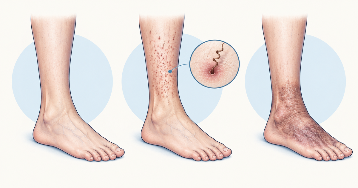
Scurvy, or vitamin C deficiency, is the collagen disease of the vitamin list, so its signs come from fragile vessels and poor wound repair. Early symptoms are lassitude, irritability, myalgia and arthralgia. After a few months come follicular hyperkeratosis, coiled hair, perifollicular haemorrhages, swollen purple gums that bleed, loose teeth and poor wound healing.
In children, pain on movement and impaired bone growth appear, and skeletal radiographs help in children but not in adults. Bleeding time, coagulation and PT are normal, which separates scurvy from vitamin K deficiency. Blood ascorbic acid below 0.6 mg/dL is marginal and below 0.2 mg/dL is deficient.
Treatment is ascorbic acid 500 to 1000 mg orally daily for 1 to 2 weeks until signs disappear, and symptoms usually resolve within that time.
Vitamin B12 Versus Folate Deficiency
Both vitamins cause a megaloblastic anaemia that looks identical on a smear, so the separation rests on the neurological examination and one biochemical test. Neurological findings can occur in B12 deficiency without any anaemia: early loss of position and vibration sense and weakness, then hyporeflexia, spasticity, extensor plantars and ataxia. Folate deficiency does not follow this pattern.
Which test separates B12 from folate deficiency? Methylmalonic acid: it is raised in B12 deficiency and normal in folate deficiency.
For the cut-offs, serum B12 below 200 pg/mL is deficient, while 200 to 350 pg/mL is borderline and needs methylmalonic acid (MMA) and homocysteine. MMA is raised in B12 deficiency and normal in folate deficiency, whereas homocysteine rises in both. Folate is low when serum folate is below 3 ng/mL, and RBC folate below 140 ng/mL is a better marker when intake has recently changed.
| Feature | Vitamin B12 deficiency | Folate deficiency |
|---|---|---|
| Megaloblastic anaemia | Yes | Yes |
| Neurological damage | Yes, can occur without anaemia | No |
| Methylmalonic acid | Raised | Normal |
| Homocysteine | Raised | Raised |
The treatment order is a safety point. MSD warns that folate supplementation can mask B12 deficiency by correcting the anaemia while neurological damage progresses, and the NHS treatment guidance says to check B12 before starting folic acid. Severe B12 deficiency is treated with 1 mg intramuscularly one to four times weekly for several weeks, then monthly. The blood count responds within about 6 weeks, while neurological recovery is slower and old deficits may be irreversible.
In pregnancy, MSD recommends folic acid 400 to 800 mcg of folic acid daily, and 4000 mcg daily after a previous neural tube defect pregnancy, starting about 3 months before conception and continuing until 12 weeks of gestation.
For the wider anaemia picture, see the anaemia classification post.
Common Mistakes With Vitamin Deficiency Diseases
Most errors come from mixing up look-alike pairs. A few habits keep them apart:
- Rickets belongs to children with growth-plate changes, and osteomalacia to adults with Looser lines.
- Scurvy has a normal PT, while vitamin K deficiency has a prolonged PT. Both bleed.
- Dry beriberi is neuropathy; wet beriberi is heart failure with warm skin.
- Pellagra and B6 deficiency can both show dermatitis, so look at the drug history and the diarrhoea and dementia.
- B12 and folate give the same smear, so use MMA and the neurological examination.
- Do not give glucose before thiamine in an at-risk patient, and do not give folate alone when B12 deficiency is possible.
A practical way to revise is to redraw both tables from memory, then check them against this page and fix the cells you missed. If a stem gives you a clue like Bitot spots, Casal necklace or perifollicular haemorrhage, name the vitamin first and the lab test second.
Putting Vitamin Deficiency Diseases Together
The vitamin deficiency diseases fall into a small set of patterns: eye and epithelium for A, bone for D, nerve and red cell for E, clotting for K, nerve and heart for thiamine, skin and gut and brain for niacin, collagen for C, and blood plus nerve for B12 and folate. Learn the signature sign for each, then the one test or rule that separates it from its nearest look-alike.
When you are ready to test yourself, work through the Biochemistry vitamins practice questions, then revisit the vitamin-derived cofactors in the Krebs cycle post. For wider practice, use the NEET PG question bank.
Practise this on Reflex
Turn what you just read into recall with 14 years of tagged PYQs.
FAQ
Frequently asked questions
The questions aspirants ask most about this topic.
Vitamins A, D, E and K are fat-soluble. The B-complex vitamins and vitamin C are water-soluble. Fat-soluble deficiency should make you think of fat malabsorption, while water-soluble deficiency points to poor diet, alcohol use disorder or interfering drugs.
Both are thiamine deficiency. Dry beriberi is a symmetric peripheral neuropathy with foot burning, calf cramps and wasting. Wet beriberi is cardiovascular, with vasodilation, tachycardia, wide pulse pressure, warm skin, oedema and heart failure.
Both cause megaloblastic anaemia. B12 deficiency can also cause neurological damage, such as loss of position and vibration sense, and it raises methylmalonic acid. In folate deficiency, methylmalonic acid is normal. Homocysteine rises in both.
In patients with alcohol use disorder or other risk of thiamine deficiency, thiamine 100 mg intravenously is given before any intravenous glucose. Glucose given first can worsen the deficiency state.
Pellagra is niacin deficiency with dermatitis, gastrointestinal symptoms such as diarrhoea, and neurological symptoms such as dementia. The rash is symmetric and photosensitive, with a pellagrous glove or boot and a Casal necklace. Maize-based diets are a typical cause.
Both can cause bleeding, but in scurvy the bleeding time, coagulation tests and PT are normal. In vitamin K deficiency the PT is prolonged and the INR raised, with a normal PTT, thrombin time, platelet count and fibrinogen.
Stay ahead in your preparation
Get expert tips, exam updates, and high-yield insights delivered straight to your inbox.
No spam. Unsubscribe anytime.
More in Subject Notes
View all → Biochemistry
Glycolysis for NEET PG: The Three Enzymes That Actually Decide the Answer
 Biochemistry
Krebs Cycle Mnemonic for NEET PG: Steps, Enzymes, and Clinical Correlations
All 8 steps in order, the rate-limiting enzymes, ATP yield, and the poisons that block specific steps.
 Anatomy
Femoral Triangle for NEET PG: Boundaries, NAVEL Contents and Femoral Hernia
Revise the femoral triangle for NEET PG: three boundaries, floor and roof, NAVEL contents, the three sheath compartments, the femoral canal and ring, and the femoral hernia link.
 Ophthalmology
Visual Pathway and Its Lesions for NEET PG: A Site-by-Site Field Defect Map
Trace the visual pathway from retina to occipital cortex and learn which lesion at each site produces which field defect, with a one-page table and worked self-test questions.