Anemia Classification for NEET PG: Morphological Types, Iron Studies, and the Differentials Examiners Actually Test
By Dr. Utsav Bhattacherjee, MBBS, MBA · 24 Aug 2026 · 9 min read
Last updated: 23 Sept 2026
On this page
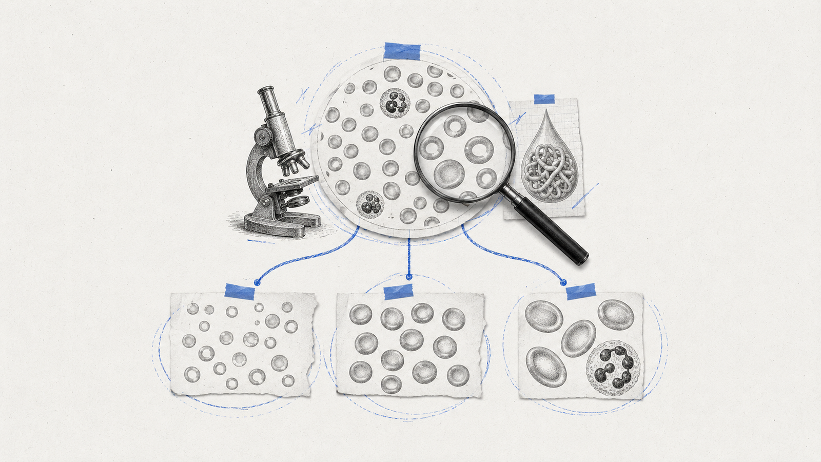
Anemia questions cut across more of the NEET PG syllabus than almost any other single topic — Medicine, Pathology, Pediatrics, and Obstetrics all lean on the same classification. Most questions don't ask you to define anemia; they hand you a hemogram or a peripheral smear and expect you to name the type on sight. That skill comes from knowing the classification cold, not from reading about anemia in general.
Morphological Classification: Start With MCV
The first sort is always by red cell size, using Mean Corpuscular Volume (MCV) from the CBC:
| Type | MCV | Common causes |
|---|---|---|
| Microcytic | Below 80 fL | Iron deficiency, thalassemia, sideroblastic anemia, anemia of chronic disease |
| Normocytic | 80–100 fL | Acute blood loss, hemolytic anemia, anemia of chronic disease (early), aplastic anemia, chronic kidney disease |
| Macrocytic | Above 100 fL | Megaloblastic anemia (B12/folate deficiency), liver disease, hypothyroidism, alcohol use, reticulocytosis |
Notice anemia of chronic disease shows up in two rows — it can present as either microcytic or normocytic depending on how established it is, which is exactly the kind of detail a well-built exam question will test.
Microcytic Anemia: The Four to Tell Apart
Iron deficiency anemia is the most common cause worldwide and the most commonly tested, but the exam rewards distinguishing it from the other three microcytic causes, not just recognizing "microcytic equals iron deficiency."
- Iron deficiency anemia (IDA): low iron stores, the most common cause globally.
- Thalassemia: an inherited defect in globin chain synthesis — disproportionately low MCV relative to the degree of anemia is a classic clue.
- Sideroblastic anemia: a defect in heme synthesis, with iron trapped inside mitochondria of developing red cells (ringed sideroblasts on bone marrow).
- Anemia of chronic disease (ACD): iron is present in the body but functionally sequestered and unavailable for erythropoiesis, typically driven by chronic inflammation.
The Iron Studies Table That Actually Separates Them
This is the single highest-yield table in the whole topic, because it's what a well-built exam question is actually testing — not "is this microcytic" but "which microcytic cause is this":
| Parameter | Iron deficiency | Anemia of chronic disease | Thalassemia |
|---|---|---|---|
| Serum iron | Low | Low | Normal |
| TIBC | High | Low | Normal |
| Ferritin | Low | Normal or high | Normal |
| Transferrin saturation | Low | Low | Normal |
The pattern worth memorizing: iron deficiency and ACD both show low serum iron, which is exactly why TIBC and ferritin are what actually separate them — TIBC rises in iron deficiency (the body is hungry for iron) and falls in ACD (inflammation suppresses transferrin production), while ferritin, an acute-phase reactant, stays normal or even rises in ACD despite the anemia. Thalassemia stands apart from both because the problem isn't iron availability at all — it's a normal amount of iron with a defective globin chain synthesis.
Macrocytic Anemia: Megaloblastic vs Non-Megaloblastic
The megaloblastic/non-megaloblastic split is the first branch point, and it's a straightforward one: megaloblastic anemia comes from impaired DNA synthesis (B12 or folate deficiency), producing large, immature-looking red cell precursors. Non-megaloblastic causes — liver disease, hypothyroidism, alcohol use, and reticulocytosis — raise MCV through entirely different mechanisms and don't share the same bone marrow picture.
B12 deficiency vs folate deficiency is the next distinction, and the exam-relevant difference is neurological: B12 deficiency can cause subacute combined degeneration of the spinal cord — a genuine neurological deficit — while folate deficiency does not. If a question pairs macrocytic anemia with neurological symptoms (paresthesias, gait disturbance, impaired proprioception), that's pointing at B12, not folate.
Normocytic Anemia: Hemolytic vs Non-Hemolytic
A normal MCV splits into two very different directions. Non-hemolytic causes include acute blood loss, aplastic anemia, chronic kidney disease, and early anemia of chronic disease. Hemolytic causes are where the exam gets more specific, because hemolytic anemia has its own classification worth knowing separately.
Hemolytic Anemia: Intrinsic vs Extrinsic
- Intrinsic (defect within the red cell itself): membrane defects (hereditary spherocytosis, hereditary elliptocytosis), enzyme defects (G6PD deficiency, pyruvate kinase deficiency), and hemoglobinopathies (sickle cell disease, thalassemia).
- Extrinsic (a normal red cell destroyed by an outside factor): autoimmune hemolytic anemia, mechanical destruction (microangiopathic hemolytic anemia, prosthetic heart valves), and infections (malaria).
Peripheral Smear Clues Worth Memorizing
A described smear finding is often the fastest route to the diagnosis in a vignette-style question:
- Target cells — thalassemia (also seen in liver disease and post-splenectomy states)
- Spherocytes — hereditary spherocytosis or autoimmune hemolytic anemia
- Sickle cells — sickle cell disease
- Schistocytes (fragmented cells) — microangiopathic hemolytic anemia, mechanical valve hemolysis
- Macro-ovalocytes with hypersegmented neutrophils — megaloblastic anemia
- Basophilic stippling — lead poisoning, thalassemia, sideroblastic anemia
Hypersegmented neutrophils specifically are worth flagging on their own — they're one of the earliest and most specific peripheral blood clues to megaloblastic anemia, sometimes appearing before the macrocytosis itself is fully established.
A Second Branch Point: Reticulocyte Count
MCV isn't the only axis worth classifying by. Reticulocyte count — corrected for the degree of anemia, since a raw retic percentage is misleading when total red cell mass has dropped — tells you whether the bone marrow is responding appropriately to the anemia or failing to compensate at all.
- Low/inappropriate reticulocyte response points to a hypoproliferative marrow: iron deficiency (once stores are truly depleted), aplastic anemia, anemia of chronic disease, or bone marrow infiltration.
- High/appropriate reticulocyte response points to the marrow working overtime to compensate: acute blood loss or hemolysis, where red cells are being destroyed or lost faster than normal but production capacity itself is intact.
This matters clinically as much as it matters for exams: a patient with hemolytic anemia and an unexpectedly low reticulocyte count is a red flag for a superimposed problem — a concurrent marrow suppression, a folate or B12 deficiency limiting the marrow's ability to respond, or an aplastic crisis (classically triggered by parvovirus B19 in a patient with an underlying hemolytic condition like sickle cell disease or hereditary spherocytosis). The exam-relevant takeaway: a hemolytic process without an elevated reticulocyte count should make you look for a second, separate problem rather than accepting the first diagnosis at face value.
Clinical Presentation Clues Worth Connecting to the Classification
Symptoms alone rarely nail the anemia type, but a few specific findings are worth linking back to mechanism rather than memorizing in isolation:
- Pica and koilonychia (spoon-shaped nails) point specifically toward iron deficiency, not anemia in general.
- Glossitis and angular stomatitis appear in both iron deficiency and B12/folate deficiency, so they don't discriminate between microcytic and macrocytic causes on their own — a reminder that not every clinical sign is diagnostic by itself.
- Jaundice with anemia should immediately raise hemolysis as a possibility, since red cell breakdown releases bilirubin — pairing this with the classification above, an elevated reticulocyte count alongside jaundice strengthens a hemolytic diagnosis considerably.
- Bone pain, particularly in children, alongside a microcytic picture, is a thalassemia clue worth knowing — ineffective erythropoiesis drives bone marrow expansion.
Anemia of chronic disease is the classic "it depends" answer that trips people up, because it can present as either microcytic or normocytic. If a question gives you a patient with a known chronic inflammatory condition (rheumatoid arthritis, chronic infection, malignancy) and a mild-to-moderate anemia without a clean fit into the classic iron-deficiency pattern, ACD deserves serious consideration even if the MCV doesn't scream "microcytic."
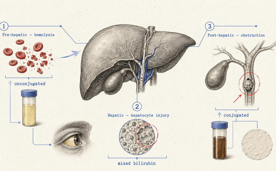
Classification-and-differential questions like this reward pattern recall built through repetition — the kind of daily, subject-wise practice Reflex's TROCAR quiz is built around. For the broader picture of how much of your NEET PG paper touches subjects like this one, see our subject-wise weightage breakdown, and for a related Medicine differential built the same way, our guide to jaundice types covers the same kind of lab-pattern recognition.
Practise this on Reflex
Turn what you just read into recall with 14 years of tagged PYQs.
FAQ
Frequently asked questions
The questions aspirants ask most about this topic.
Sorting by MCV into microcytic (under 80 fL), normocytic (80–100 fL), or macrocytic (over 100 fL) — this determines which set of causes to consider next.
Both show low serum iron, but TIBC and ferritin diverge: TIBC is high and ferritin is low in iron deficiency, while TIBC is low and ferritin is normal-to-high in anemia of chronic disease.
Both cause megaloblastic, macrocytic anemia, but only B12 deficiency causes neurological involvement, including subacute combined degeneration of the spinal cord.
Thalassemia, sideroblastic anemia, and anemia of chronic disease — iron studies are what separate these from each other.
Macro-ovalocytes and hypersegmented neutrophils — the hypersegmented neutrophils in particular can appear before macrocytosis is fully established.
No — it can present as either microcytic or normocytic depending on how long-standing the underlying inflammatory condition is, which is a common source of confusion in exam questions.
About the author
Stay ahead in your preparation
Get expert tips, exam updates, and high-yield insights delivered straight to your inbox.
No spam. Unsubscribe anytime.
More in Medicine
View all → Medicine
Nephrotic Syndrome vs Nephritic Syndrome: The Proteinuria Threshold That Explains Everything Else
 Medicine
Murmur Grading for NEET PG: The Levine Scale, and Why Louder Doesn't Always Mean Worse
 Medicine
Jaundice Types for NEET PG: Pre-Hepatic, Hepatic, and Post-Hepatic Classification
Jaundice classification for NEET PG — pre-hepatic, hepatic, and post-hepatic types, the lab pattern that tells them apart, and Courvoisier's law explained.
 Medicine
Clubbing Causes for NEET PG: Grades, Schamroth’s Sign, and the Full List
The four grades, Schamroth’s window test, and the complete pulmonary, cardiac and GI cause list.