Nephrotic Syndrome vs Nephritic Syndrome: The Proteinuria Threshold That Explains Everything Else
By Dr. Utsav Bhattacherjee, MBBS, MBA · 3 Sept 2026 · 12 min read
Last updated: 23 Sept 2026
On this page
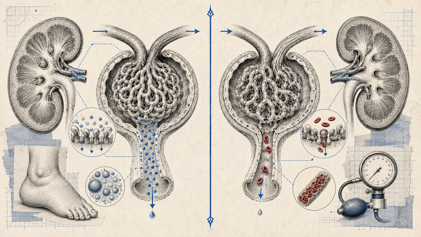
Nephrotic and nephritic syndrome get confused constantly because both names sound similar and both involve the kidney's filtering units — but they're distinguished by one core mechanism: how much protein is leaking through the glomerulus, and whether that leak is inflammatory or not. Get that mechanism straight, and the entire cluster of findings for each syndrome follows logically rather than needing to be memorized as an arbitrary list.
The Core Distinction
Nephrotic syndrome is fundamentally a non-inflammatory problem — damage to the podocytes and glomerular basement membrane that lets massive amounts of protein leak into the urine, without significant inflammatory cell infiltration or bleeding. Nephritic syndrome is the opposite: a genuinely inflammatory process — glomerulonephritis — where immune-mediated injury to the glomerulus lets red blood cells escape into the urine and impairs the kidney's filtering function enough to cause fluid retention and hypertension. Protein loss happens in both, but it's the defining, massive feature of nephrotic syndrome and a secondary, more modest feature of nephritic syndrome.
Nephrotic Syndrome: The Full Clinical Picture
The defining feature is massive proteinuria — conventionally set at greater than 3.5 grams per day in adults — and everything else in the syndrome follows from the consequences of losing that much protein. Hypoalbuminemia develops because the liver can't synthesize albumin fast enough to keep up with urinary losses. That falling albumin drops plasma oncotic pressure, which is what actually drives the edema — fluid shifts out of the vasculature into the tissues because there's less oncotic force pulling it back in, classically producing periorbital edema on waking and progressing to more generalized swelling through the day.
Hyperlipidemia rounds out the classic tetrad, and it's a detail worth understanding rather than just memorizing: the liver, working overtime to compensate for albumin losses, ends up non-specifically ramping up synthesis of other proteins too, including lipoproteins — so cholesterol and triglycerides both rise as a side effect of the liver's attempt to fix the albumin problem, not as an independent process.
A less commonly emphasized but genuinely important consequence: nephrotic syndrome is a hypercoagulable state. Antithrombin III, a natural anticoagulant protein, is lost in the urine alongside albumin, tipping the balance toward clotting — renal vein thrombosis is a recognized complication specifically associated with nephrotic syndrome, and a vignette describing sudden flank pain and hematuria in a patient with known nephrotic syndrome is testing exactly this connection.
Nephritic Syndrome: The Full Clinical Picture
The defining feature is hematuria, classically described as "smoky" or "tea-colored" urine, and the single most specific finding on urinalysis is red blood cell casts — their presence essentially confirms the bleeding is originating in the glomerulus itself, not lower in the urinary tract, since casts can only form within the nephron's tubules.
The inflammatory damage to the glomerulus also impairs filtration, which produces the rest of the picture: oliguria (reduced urine output, as the filtering capacity itself is compromised) and hypertension (from fluid retention as filtration falls, activating the renin-angiotensin system further). Proteinuria is present too, but it's generally sub-nephrotic — well under the 3.5g/day threshold — which is the key quantitative distinction from nephrotic syndrome even though both syndromes technically involve protein in the urine.
Side by Side
| Nephrotic Syndrome | Nephritic Syndrome | |
|---|---|---|
| Core mechanism | Non-inflammatory podocyte/GBM damage | Inflammatory glomerulonephritis |
| Proteinuria | Massive (>3.5g/day) | Mild-moderate (<3.5g/day) |
| Hematuria | Absent or minimal | Prominent, with RBC casts |
| Edema | Prominent, from hypoalbuminemia | Present, from fluid retention |
| Blood pressure | Usually normal | Elevated |
| Hyperlipidemia | Present | Absent |
Causes of Nephrotic Syndrome
Minimal change disease is the most common cause in children, and its name describes exactly what you'd see (or rather, wouldn't see) on light microscopy — the glomerulus looks essentially normal, with the damage only visible on electron microscopy as effacement (flattening) of the podocyte foot processes. It's notable for being strongly steroid-responsive, which is itself a useful diagnostic and prognostic clue in a pediatric nephrotic syndrome vignette.
Focal segmental glomerulosclerosis (FSGS) is a leading cause in adults, particularly in populations of African descent, and unlike minimal change disease, it shows genuine sclerotic (scarring) changes on biopsy, affecting only some glomeruli (focal) and only part of each affected glomerulus (segmental) — the name is literally describing the biopsy pattern.
Membranous nephropathy is the classic cause in older adults, and it's worth remembering as frequently secondary to something else — malignancy, hepatitis B, or autoimmune disease being the recognized associations — which makes a new membranous nephropathy diagnosis in an older adult a reasonable trigger for age-appropriate cancer screening, not just kidney-focused workup.
Diabetic nephropathy deserves separate mention as the single most common cause of nephrotic syndrome overall once you account for the sheer prevalence of diabetes — the classic biopsy finding is Kimmelstiel-Wilson nodules, nodular glomerulosclerosis that's specific enough to the condition that its description alone usually settles the diagnosis in a vignette.
Causes of Nephritic Syndrome
Post-streptococcal glomerulonephritis is the classic cause to know cold: it follows a group A streptococcal throat or skin infection, but critically, with a latent period of 1–3 weeks between the infection and the onset of kidney symptoms — this delay reflects the time needed for immune complexes to form and deposit in the glomerulus, and it's the detail that separates this diagnosis from the next one.
IgA nephropathy (Berger's disease) is actually the most common cause of glomerulonephritis worldwide, and the timing is the opposite of post-streptococcal GN: it classically presents with gross hematuria within 1–2 days of an upper respiratory or gastrointestinal infection — essentially concurrent with the infection rather than weeks after it. This rapid-onset-versus-delayed-onset contrast against post-streptococcal GN is one of the most reliably tested distinctions in this whole topic.
Rapidly progressive glomerulonephritis (RPGN) is the most aggressive presentation, characterized by loss of renal function over days to weeks rather than the more gradual course of other causes. On biopsy, it's defined by crescents — crescent-shaped proliferations within Bowman's space — and it can result from several different underlying triggers (anti-GBM disease, immune complex disease, or ANCA-associated vasculitis), unified by this shared aggressive histological pattern rather than one single cause.
Lupus nephritis deserves a specific mention because systemic lupus erythematosus is genuinely unusual in being able to produce either nephritic or nephrotic patterns, or occasionally both together, depending on which histological class of lupus nephritis is present — a reminder that the nephrotic/nephritic split is a description of the clinical picture, not a rigid categorization every kidney disease fits cleanly into.
When the Two Overlap
Membranoproliferative glomerulonephritis (MPGN) is worth knowing specifically because it can present with features of both syndromes simultaneously — nephrotic-range proteinuria alongside nephritic-pattern hematuria and RBC casts. This isn't a contradiction of the framework above so much as a reminder that nephrotic and nephritic are two ends of a spectrum of glomerular injury, and a small number of conditions genuinely straddle both ends rather than sitting cleanly on one side.
How Proteinuria Is Actually Quantified
The 3.5g/day threshold sounds precise, but it's worth knowing how that number actually gets measured in practice, since the gold-standard method and the practical everyday method aren't the same test. A 24-hour urine collection is the traditional gold standard — every drop of urine over a full day is collected and the total protein measured directly — but it's genuinely burdensome for patients to complete accurately and slow to turn around, which limits how often it's actually used as a first-line test.
In practice, a spot urine protein-to-creatinine ratio has become the far more commonly used surrogate — a single urine sample, measuring the ratio of protein to creatinine, which correlates well enough with 24-hour protein excretion to substitute for it in most clinical situations without requiring a full day of collection. A ratio above roughly 3.5 (in the same units this test is conventionally reported in) is generally taken as consistent with nephrotic-range proteinuria, mirroring the 3.5g/day threshold from the full collection method.
This distinction matters for reading a vignette correctly: a spot ratio and a 24-hour total are reported differently and shouldn't be confused with each other, even though both are ultimately being used to answer the same underlying question — is this patient losing protein at a nephrotic-range rate or not.
For the same pattern-recognition approach applied to a different lab-driven diagnosis, see our guide to anemia classification, and for how much of the paper Medicine carries overall, our subject-wise weightage breakdown.
Practise this on Reflex
Turn what you just read into recall with 14 years of tagged PYQs.
FAQ
Frequently asked questions
The questions aspirants ask most about this topic.
Nephrotic syndrome is a non-inflammatory process causing massive proteinuria (>3.5g/day), hypoalbuminemia, edema, and hyperlipidemia. Nephritic syndrome is an inflammatory process (glomerulonephritis) causing hematuria with RBC casts, hypertension, and only mild-to-moderate proteinuria.
The liver increases protein synthesis to compensate for urinary albumin loss, but this compensation isn't specific to albumin — it non-specifically increases lipoprotein synthesis too, raising cholesterol and triglycerides as a side effect.
Minimal change disease, which looks essentially normal on light microscopy and only shows podocyte foot process effacement on electron microscopy. It's strongly steroid-responsive, which is itself a useful clinical clue.
Timing relative to the preceding infection. Post-streptococcal GN has a latent period of 1–3 weeks after a strep infection. IgA nephropathy presents with gross hematuria within 1–2 days of a respiratory or GI infection.
Antithrombin III, a natural anticoagulant, is lost in the urine along with albumin, tipping the body toward a hypercoagulable state. Renal vein thrombosis is a recognized complication specifically linked to this mechanism.
About the author
Stay ahead in your preparation
Get expert tips, exam updates, and high-yield insights delivered straight to your inbox.
No spam. Unsubscribe anytime.
More in Medicine
View all → Medicine
Murmur Grading for NEET PG: The Levine Scale, and Why Louder Doesn't Always Mean Worse
 Medicine
Anemia Classification for NEET PG: Morphological Types, Iron Studies, and the Differentials Examiners Actually Test
Anemia questions cut across Medicine, Pathology, Pediatrics and Obstetrics, and most of them hand you a hemogram and expect you to name the type on sight. The morphological classification, the iron studies table, and the smear clues that separate the differentials.
 Medicine
Jaundice Types for NEET PG: Pre-Hepatic, Hepatic, and Post-Hepatic Classification
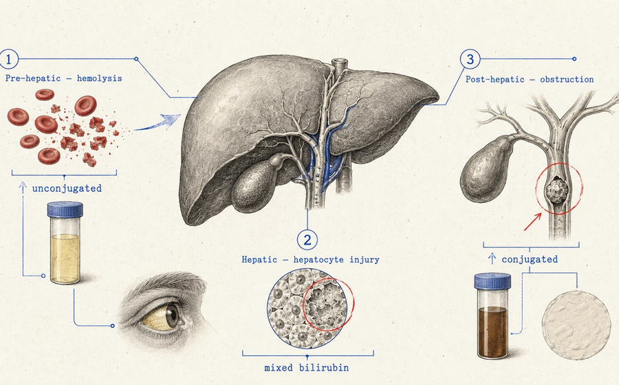
Jaundice classification for NEET PG — pre-hepatic, hepatic, and post-hepatic types, the lab pattern that tells them apart, and Courvoisier's law explained.
 Medicine
Clubbing Causes for NEET PG: Grades, Schamroth’s Sign, and the Full List
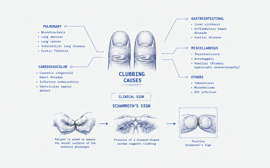
The four grades, Schamroth’s window test, and the complete pulmonary, cardiac and GI cause list.