Colles Fracture for NEET PG: Dinner Fork Deformity, Smith and Barton Compared, and the Complications
Reflex · 5 Nov 2026 · 10 min read
Last updated: 5 Nov 2026
On this page
A Colles fracture is a distal radius fracture in which the broken fragment tilts toward the back of the wrist. Three details separate it from its look-alikes: the direction of angulation, whether the joint surface is involved, and the way the patient fell. Once you hold those three in mind, Colles, Smith and Barton stop blurring together.
This post builds the comparison step by step. It covers mechanism, the dinner fork deformity, radiographic features, management by age, and the complications you should be able to list without hesitation. For a wider view of how fractures are described, see our guide to types of fractures.
Key Takeaways
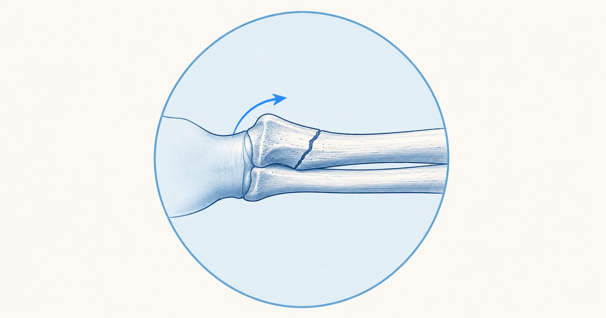
A Colles fracture breaks the distal radial metaphysis without entering the joint (extra-articular), and the distal fragment is angulated dorsally and impacted. The usual cause is a FOOSH, a fall on an outstretched hand that leaves the wrist extended.
A Smith fracture is the mirror image: the distal fragment tilts volarly (toward the palm). It is called a "reverse Colles".
A Barton fracture is different in kind. It is intra-articular and usually comes with radiocarpal subluxation or dislocation; the dorsal type resembles Colles and the volar type is the reverse Barton.
AAOS guidance for people under about 65: surgery helps when, after reduction, radial shortening exceeds 3 mm, dorsal tilt exceeds 10 degrees, or an articular step-off exceeds 2 mm.
In adults aged about 65 and over, surgery does not improve long-term patient-reported outcomes compared with non-operative care.
Key complications: malunion (dinner fork deformity), median nerve compression and carpal tunnel syndrome, CRPS, wrist stiffness, secondary osteoarthritis and late extensor pollicis longus (EPL) tendon rupture.
What Is a Colles Fracture?
In a Colles fracture, the break lies in the distal radial metaphysis, outside the joint (extra-articular), and the distal fragment is both dorsally angulated and impacted. Radiopaedia describes it as not involving the articular surface. That single point matters, because it is what separates it from Barton fractures later in this post.
The term sits inside the broader family of distal radius fractures, which also includes Smith, Barton and styloid injuries. Learn the strict definition first: dorsal angulation, impaction and an intact joint surface.
Mechanism and Who Gets a Colles Fracture
The classic mechanism is a fall on an outstretched hand with the forearm pronated and the wrist extended. According to Radiopaedia, the proximal carpal row, mainly the lunate and scaphoid, drives the radius dorsally and along its long axis. The result is dorsal angulation plus impaction.
Age changes the story. AAOS OrthoInfo notes that people with osteoporosis can break the wrist in a fall from standing height. Younger people usually need a high-force event such as a road crash or a fall from a ladder or bicycle.
This has a practical consequence. A fragility wrist fracture in an older adult can be the first sign of osteoporosis, so it is worth thinking about bone health as well as the wrist.
Dinner Fork Deformity: Why the Wrist Looks Like a Fork
The dinner fork deformity is the dorsal displacement of the carpal bones and distal fragment, which gives the wrist a bent, fork-like profile in side view. Picture the back of the hand rising into a hump just beyond the wrist, with a dip proximal to it. The shape follows directly from the dorsal angulation.
Typical clinical findings are pain, swelling, bruising and tenderness over the distal radius. Numb fingers suggest nerve compression, and any open wound must be excluded. Acute carpal tunnel syndrome and compartment syndrome are uncommon but urgent, so check sensation and the tightness of the forearm early. Our post on compartment syndrome explains why late signs should never be awaited.
The same deformity can appear later if the fracture heals in a bad position. In that case it is a complication (malunion), not an acute finding.
Radiological Features of a Colles Fracture
Radiopaedia lists the following on plain films: dorsal angulation, impaction and radial shortening, visible on the lateral and AP views. A pronator quadratus sign is generally seen, and the ulnar styloid is fractured in up to half of cases. CT is added if intra-articular extension is suspected.
Use this as a short reading routine on a wrist film:
- On the lateral view, decide which way the distal fragment tilts.
- On the AP view, look for impaction and loss of radial length.
- Look at the joint surface for any step-off.
- Check the ulnar styloid.
If the lateral view shows dorsal tilt with an intact joint surface, think Colles. If the tilt is volar, think Smith.
Colles vs Smith vs Barton: Comparison Table
The table below puts the three eponyms side by side. Information comes from Radiopaedia entries on Colles, Smith, Barton and reverse Barton fractures.
| Feature | Colles | Smith | Barton |
|---|---|---|---|
| Direction of distal fragment | Dorsal angulation | Volar (palmar) angulation | Dorsal type: dorsal; volar type (reverse Barton): volar displacement |
| Joint surface | Extra-articular | Classically extra-articular | Intra-articular |
| Usual mechanism | Fall on outstretched hand, wrist extended | Fall onto a flexed wrist, or a direct blow to the dorsum of the wrist | Not covered here |
| Deformity | Dinner fork | Garden spade (after malunion) | Dorsal subluxation or dislocation of the radiocarpal joint in the dorsal type |
| Other name | None | Reverse Colles | Reverse Barton for the volar type |
| Typical associated problem | Median nerve compression, malunion | Carpal tunnel syndrome from volar displacement | Radiocarpal joint disruption |
Two cautions apply. First, the term Smith fracture is sometimes stretched to cover intra-articular fractures with volar displacement and juxta-articular fractures. Radiopaedia calls the reverse Barton fracture a "type II Smith fracture", which is oblique and intra-articular.
Second, the Barton fracture has a clear definition: a distal radius fracture that extends through the dorsal aspect to the articular surface but not the volar aspect, usually with dorsal subluxation or dislocation of the radiocarpal joint. It is named after John Rhea Barton. Barton fracture treatment is outside the scope of this post.
A Memory Aid for the Direction of Tilt
Here is an original memory aid (not from any source): Colles Climbs to the back, Smith Sags to the palm. It fixes the direction of the distal fragment in one line. Add one extra rule: Barton always means the joint surface is involved.
Management of Colles Fracture
Management depends on stability, joint involvement and age. The first step is reduction and immobilisation, with surgery for fractures that are unstable or fail to hold their position.
Non-Operative Treatment
Radiopaedia describes closed reduction followed by a cast from below the elbow to the metacarpal heads. Traditionally the wrist is held flexed and in ulnar deviation. According to OrthoInfo, a splint often goes on first so that swelling can settle, and a cast follows later. The cast is typically removed at around 4 to 6 weeks, after which physical therapy begins.
Smith fractures are handled similarly: most are closed-reduced and casted, with open reduction and internal fixation (ORIF) usually needed when the fracture cannot be reduced or is unstable after reduction.
When to Operate: AAOS Thresholds
The AAOS and ASSH clinical practice guideline fact sheet (December 2020) gives age-based advice. Learn it as a two-branch decision.
| Patient group | AAOS 2020 guidance |
|---|---|
| Non-geriatric (generally under 65) | Surgery improves radiographic and patient-reported outcomes when, after reduction, there is radial shortening over 3 mm, dorsal tilt over 10 degrees, or intra-articular step-off or displacement over 2 mm |
| Older adults (generally 65 and over) | Surgery does not improve long-term patient-reported outcomes compared with non-operative treatment |
These numbers are the AAOS values. Some secondary sources quote a different shortening cut-off, such as 5 mm, so use the AAOS figures as your reference. A useful way to apply them is to measure the post-reduction film, not the initial one.
Fixation Options
For complete articular or unstable fractures, the AAOS fact sheet reports that fixation techniques did not differ in major outcomes. Volar locking plates, however, speed functional recovery at about three months. OrthoInfo lists plates, screws, pins and external fixators as the available tools.
Complications of Colles Fracture
The complication list is best sorted by timing and by structure. Radiopaedia names the main ones, and AAOS OrthoInfo adds the practical points about pain and stiffness.
| Complication | Key points |
|---|---|
| Malunion | Leads to dinner fork deformity; can cause pain, weak grip and reduced forearm rotation |
| Median nerve compression | Median nerve palsy and post-traumatic carpal tunnel syndrome |
| CRPS (reflex sympathetic dystrophy) | Persistent severe pain can signal it |
| Wrist stiffness | Common; improves for up to 2 years; more likely after severe injuries or in people over 50 |
| Secondary osteoarthritis | More likely with intra-articular involvement |
| EPL tendon rupture | A late complication of the distal radius fracture |
| Nonunion | Rare |
Incidence figures for these complications vary and are not given here, so the table stays qualitative. Learn the list, then attach the clinical clue to each item.
Nerve and Tendon Problems
Median nerve compression is the neurological complication to know best. It can present acutely or later as carpal tunnel syndrome. Extensor pollicis longus (EPL) rupture is a recognised late complication of distal radius fracture.
Pain, Stiffness and Joint Damage
Persistent severe pain should raise the question of CRPS. Stiffness is common and keeps improving for up to two years. Secondary osteoarthritis is more likely when the fracture reached the joint surface, and the AAOS threshold for an articular step-off is 2 mm.
Common Mistakes When Comparing Colles, Smith and Barton Fractures
These are mix-ups that are easy to make while revising, so it helps to name them.
- Naming by deformity alone. The dinner fork shape fits dorsal displacement, but a Barton dorsal type can also shift the carpus dorsally. Check whether the joint surface is involved.
- Treating Barton as "just another Colles". Radiopaedia notes a similarity to Colles in the dorsal type, but the fracture is intra-articular and the radiocarpal joint is disrupted.
- Using a single age-blind rule for surgery. The AAOS guidance splits at about 65 years, and the outcome benefit of surgery in older adults is not shown.
- Quoting the wrong threshold. The set of 3 mm shortening, 10 degrees dorsal tilt and 2 mm step-off belongs together as the AAOS values.
- Forgetting the bone health angle. A wrist fracture from a standing-height fall in an older adult points toward osteoporosis.
To practise sorting these on clinical stems, work through the upper limb trauma practice questions. Fracture patterns in children are a separate system, covered in our Salter-Harris classification post.
Colles Fracture Summary: Three Questions
Think of a Colles fracture as a three-question problem: which way does the fragment tilt, is the joint surface involved, and how old is the patient? Dorsal tilt with an intact joint is Colles, volar tilt is Smith, and a joint-involving fracture is Barton. Management follows the same logic, with AAOS thresholds guiding surgery in people under about 65 and non-operative care being a reasonable approach in older adults. Complications such as malunion, median nerve compression, CRPS, stiffness, osteoarthritis and EPL rupture complete the picture.
Practise this on Reflex
Turn what you just read into recall with 14 years of tagged PYQs.
FAQ
Frequently asked questions
The questions aspirants ask most about this topic.
A Colles fracture is an extra-articular fracture of the distal radial metaphysis with dorsal angulation and impaction of the distal fragment. Most cases follow a FOOSH (fall on an outstretched hand), in which the wrist is extended on impact.
They differ in which way the distal fragment tilts. A Colles fracture angulates dorsally, whereas a Smith fracture angulates volarly, toward the palm, which is why it is called a reverse Colles. Smith fractures usually result from a fall onto a flexed wrist or a blow to the back of the wrist.
Dorsal displacement of the distal fragment and carpal bones gives the wrist a bent profile in side view, like the curve of a dinner fork. It can also develop later if the fracture heals in a malunited position.
For non-geriatric patients, generally under 65, AAOS guidance supports surgery when, after reduction, radial shortening is over 3 mm, dorsal tilt is over 10 degrees, or intra-articular step-off or displacement is over 2 mm. In adults generally 65 and over, surgery does not improve long-term patient-reported outcomes compared with non-operative treatment.
The main ones are malunion with dinner fork deformity, median nerve compression and carpal tunnel syndrome, complex regional pain syndrome, wrist stiffness, secondary osteoarthritis and late extensor pollicis longus tendon rupture. Nonunion is rare.
Stay ahead in your preparation
Get expert tips, exam updates, and high-yield insights delivered straight to your inbox.
No spam. Unsubscribe anytime.
More in Subject Notes
View all → Orthopedics
Types of Fractures for NEET PG: The Two Axes Every Question Actually Tests
 Orthopedics
Salter-Harris Classification for NEET PG: The SALTR Mnemonic, Explained Properly
Salter-Harris questions test two things: reading the type off an X-ray description, and knowing which type carries the worst prognosis. The SALTR mnemonic, why Type V is the trap, and how management differs across the five types.
 Anesthesia
ASA Physical Status Classification for NEET PG: Classes I to VI, Examples and the E Modifier
A clear walk through the ASA physical status classification: what each class means, example patients, the E modifier for emergencies, and the sorting mistakes to avoid.
 Psychiatry
Defense Mechanisms for NEET PG: Mature, Immature and Neurotic Defenses with One Example Each
Learn defense mechanisms as a ladder from pathological to mature, with one worked example per level, clear notes on projection and sublimation, and where Vaillant's scheme and the DMRS disagree.