Compartment Syndrome for NEET PG: Why Pulselessness Is the Sign That Comes Last, Not First
By Dr. Utsav Bhattacherjee, MBBS, MBA · 21 Sept 2026 · 13 min read
Last updated: 23 Sept 2026
On this page
Compartment syndrome questions are built around one critical trap: pulselessness feels like it should be the defining sign, but it's actually one of the latest and least reliable findings. The sign that actually matters for early diagnosis is pain that doesn't match the injury, and missing that distinction is exactly what costs limbs
The Mechanism
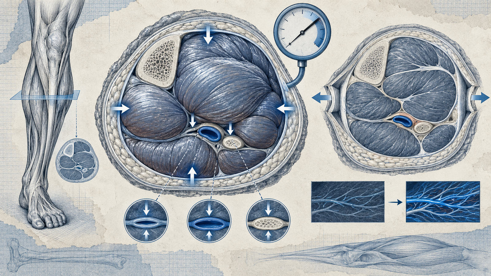
Compartment syndrome develops when pressure inside a closed fascial compartment rises high enough to compromise capillary perfusion to the muscles and nerves within it. Fascia doesn't stretch to accommodate swelling, so as pressure builds — from bleeding, edema following a fracture, or reperfusion after a period of ischemia — the compartment becomes a closed space fighting its own contents for space. Once tissue pressure approaches diastolic blood pressure closely enough, capillary flow into the compartment stops even though a major artery further upstream may still have a palpable pulse.
This is the single most important conceptual point in the entire topic: compartment syndrome is a problem of the microcirculation within the compartment, not necessarily the major vessels supplying the limb, which is exactly why distal pulses can remain normal well into the process.
Why Pain Out of Proportion Comes First
Nerve tissue is exquisitely sensitive to reduced perfusion, more so than muscle, which is why pain is the earliest reliable symptom — specifically, pain that is disproportionate to the apparent severity of the injury, and pain that worsens dramatically with passive stretch of the muscles in the affected compartment. A patient with a seemingly stable fracture who reports pain far beyond what the injury should produce, especially pain that intensifies when you passively extend their fingers or toes, is describing compartment syndrome until proven otherwise — this single finding should trigger urgent action well before any of the other classic signs appear.
The Six P's, and Why Their Order Matters
Pain and paresthesia are early findings, reflecting the nerve tissue's sensitivity to reduced perfusion. Pallor and poikilothermia (coolness) develop as perfusion continues to fall. Pulselessness and paralysis are late findings, appearing only once the compartment pressure has risen high enough to compromise larger vessels and cause frank nerve or muscle infarction — by the time pulselessness appears, irreversible muscle and nerve damage may already be well underway. Waiting for a pulse to disappear before diagnosing compartment syndrome means diagnosing it dangerously late.
Common Causes Worth Recognizing
Fractures are the leading cause, particularly of the tibia and the distal radius, with bleeding and swelling within the fascial compartment following the injury itself. Crush injuries produce compartment syndrome through direct tissue damage and subsequent swelling. Reperfusion injury after a period of vascular occlusion — restoring blood flow to a limb after prolonged ischemia — can paradoxically trigger compartment syndrome as the previously ischemic tissue swells dramatically once blood flow resumes. Tight casts or dressings can also produce a compartment-syndrome-like picture by external constriction rather than internal swelling, which is exactly why a patient in a cast reporting disproportionate pain needs the cast evaluated, not just more analgesia.
How the Diagnosis Is Actually Confirmed
While the clinical picture — disproportionate pain, pain on passive stretch — is often enough to prompt urgent action, compartment pressure can be measured directly using a needle manometer inserted into the suspected compartment. A measured pressure within 30mmHg of the patient's diastolic blood pressure is generally considered diagnostic, since this narrow gap between compartment pressure and diastolic pressure is what actually determines whether capillary perfusion can be sustained. In practice, when the clinical suspicion is strong, treatment is not delayed to await pressure measurement confirmation, since the entire point of early diagnosis is to intervene before irreversible damage accumulates.
Treatment: Fasciotomy
The definitive treatment is emergency fasciotomy — surgically incising the fascia to release the pressure within the compartment, restoring perfusion before permanent damage occurs. This is a time-critical intervention; delay converts a reversible perfusion problem into permanent muscle necrosis and nerve damage. Any external constricting factor (a tight cast, dressing, or splint) should be released immediately as a first step while arranging for definitive fasciotomy, since removing an external cause costs nothing and may partially relieve pressure while surgical intervention is arranged.
The Complication Worth Knowing by Name: Volkmann's Contracture
Volkmann's ischemic contracture is the classic long-term complication of untreated or delayed compartment syndrome, classically described in the forearm following a supracondylar humerus fracture in children. Muscle tissue that has undergone ischemic necrosis is replaced by fibrous scar tissue, which contracts over time and produces a fixed flexion deformity of the wrist and fingers — a permanent, disabling consequence of a problem that was, at the point of the missed pain-out-of-proportion sign, still entirely reversible.
The Leg: Four Compartments Worth Knowing
The lower leg is divided into four fascial compartments, and knowing which nerve and function each contains helps predict the specific deficit compartment syndrome in that location would produce. The anterior compartment contains the deep peroneal nerve and the muscles responsible for dorsiflexion, so anterior compartment syndrome classically produces a foot drop and sensory loss in the first web space. The deep posterior compartment contains the tibial nerve and the muscles responsible for toe flexion, so involvement there affects plantar sensation and toe flexion specifically.
Why Analgesia Requirements Are Themselves a Clinical Clue
A genuinely useful bedside pattern, worth holding onto alongside the formal signs: a patient whose pain requirements are escalating faster than the injury or the postoperative course would typically justify, particularly pain that isn't responding to doses that would normally be adequate, deserves specific reassessment for compartment syndrome rather than simply escalating the analgesic dose further. Treating the pain as the problem to solve, rather than as a signal pointing toward an underlying process, is a recognized way this diagnosis gets missed or delayed in practice — the pain is data, not just a symptom to suppress.
This is particularly relevant in the postoperative or post-fracture setting, where regional nerve blocks or patient-controlled analgesia can mask the early pain signal that would otherwise prompt earlier suspicion. A block that seems to be working unusually poorly, or a PCA usage pattern climbing steeply, is worth treating as a possible red flag rather than simply a sign the initial pain control plan needs adjusting.
Reading a compartment syndrome vignette efficiently means weighting the pain description heavily above every other finding offered: a normal pulse or normal color in the described limb should never be read as reassurance against the diagnosis, since both are expected to be normal at exactly the stage when intervention still has the best chance of preventing permanent damage.
Acute vs Chronic Exertional Compartment Syndrome
Everything discussed so far describes acute compartment syndrome, a true surgical emergency. A separate, distinct entity worth not confusing with it is chronic exertional compartment syndrome, seen in athletes and characterized by a recurring, predictable pattern: pain and tightness in a specific compartment that builds during a consistent level of exertion (often running) and resolves reliably with rest, only to recur at a similar exertion threshold each time. Unlike acute compartment syndrome, this is not a surgical emergency in the moment it's presenting — it's investigated electively with pressure measurements taken before and after a provocative exercise test, and while fasciotomy can be a treatment option for confirmed chronic exertional compartment syndrome, the urgency and clinical approach are entirely different from the acute presentation this guide otherwise focuses on.
Distinguishing the two by history alone is usually straightforward: a single traumatic event or fracture followed by escalating, non-resolving pain points toward the acute emergency; a recurring pattern tied to a consistent activity threshold, resolving with rest each time, points toward the chronic exertional form instead.
For the same emergency-recognition approach applied to a different surgical topic, see our guide to types of shock, and for how much of the paper Surgery carries overall, our subject-wise weightage breakdown.
Practise this on Reflex
Turn what you just read into recall with 14 years of tagged PYQs.
FAQ
Frequently asked questions
The questions aspirants ask most about this topic.
Compartment syndrome compromises capillary perfusion within the compartment, not necessarily the major artery supplying the limb. Tissue pressure can rise enough to stop capillary flow while a pulse remains palpable further downstream.
Pain out of proportion to the injury, and pain that worsens significantly with passive stretch of the muscles in the affected compartment — both appear well before pallor, pulselessness, or paralysis.
By the time compartment pressure rises high enough to compromise a palpable pulse, significant and potentially irreversible muscle and nerve damage may already have occurred.
A measured compartment pressure within 30mmHg of the patient's diastolic blood pressure. This narrow gap is what determines whether capillary perfusion within the compartment can be sustained.
A permanent flexion deformity of the wrist and fingers resulting from untreated compartment syndrome, classically following a supracondylar humerus fracture in children. Ischemic muscle necrosis is replaced by contracting fibrous scar tissue.
The entire value of early diagnosis is intervening before irreversible muscle and nerve damage accumulates. When clinical suspicion is strong, delaying treatment to confirm with pressure measurement defeats that purpose.
About the author
Stay ahead in your preparation
Get expert tips, exam updates, and high-yield insights delivered straight to your inbox.
No spam. Unsubscribe anytime.
More in Subject Notes
View all → Surgery
Burns for NEET PG: The Rule of Nines, Burn Depth and the Parkland Formula Worked Step by Step
 Surgery
Hernia Types and Classification for NEET PG
Direct vs indirect inguinal hernia, Hesselbach’s triangle, and why femoral hernias strangulate most.
 Radiology
FAST Scan for NEET PG: The Four Windows, eFAST, and What a Positive Result Means
 Dermatology