Femoral Triangle for NEET PG: Boundaries, NAVEL Contents and Femoral Hernia
Reflex · 2 Nov 2026 · 9 min read
Last updated: 2 Nov 2026
On this page
The femoral triangle is a small space at the top of the front of the thigh, and it carries the main vessels and nerve of the lower limb. For revision, cover four things: its boundaries, the order of its contents, what lies inside the femoral sheath, and why a femoral hernia strangulates so easily. This post covers all four in the order you would need them in an exam, with the common traps flagged.
The femoral triangle is also called Scarpa's triangle (Australas J Ultrasound Med, 2024). Learn both names.
Key Takeaways
Boundaries: inguinal ligament (superior, the base), medial edge of sartorius (lateral), medial edge of adductor longus (medial). Mnemonic SAIL.
Floor: iliopsoas laterally and pectineus medially. Roof: skin, subcutaneous tissue, an extension of Scarpa's fascia, the great saphenous vein, superficial lymph nodes and fascia lata.
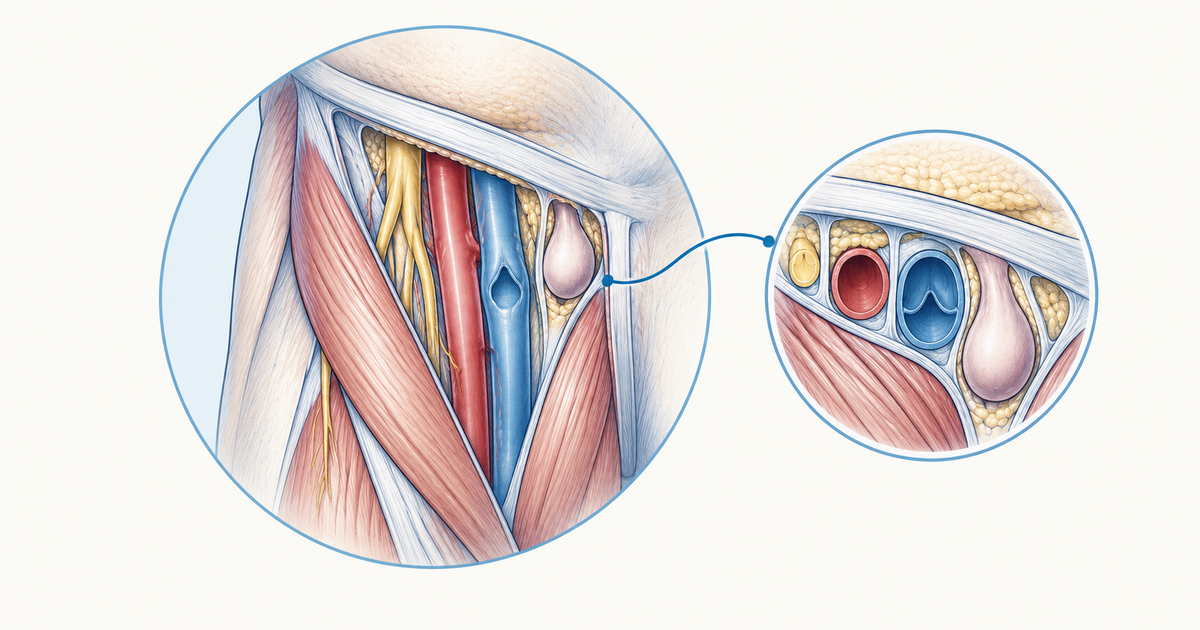
The memory aid NAVEL covers the contents: the femoral Nerve sits most laterally, followed by the Artery, the Vein, the Empty space that is the femoral canal, and the Lymphatics medially.
The femoral sheath has three compartments. The femoral branch of the genitofemoral nerve shares the lateral compartment with the artery, the vein has the intermediate one, and the medial one is the femoral canal. The femoral nerve is not enclosed: it runs outside the sheath, on its lateral side.
Strangulation risk is high because the femoral ring has rigid borders. This ring, the opening at the top of the canal, is where femoral hernias start.
A femoral hernia stays lateral to the pubic tubercle, which helps separate it from an inguinal hernia.
Boundaries of the femoral triangle
The femoral triangle has three boundaries: the inguinal ligament forms the base, the medial edge of sartorius forms the lateral border, and the medial edge of adductor longus forms the medial border (Radiopaedia: Femoral triangle; PMC11671740). The base is on top and the triangle points downward.
| Boundary | Structure |
|---|---|
| Superior (base) | Inguinal ligament |
| Lateral | Medial edge of sartorius |
| Medial | Medial edge of adductor longus |
A memory aid, not a textbook fact: SAIL stands for Sartorius, Adductor longus, Inguinal Ligament. It lists the three borders but does not tell you which is which, so learn the table too.
Where the triangle leads
Distally, the femoral vessels and the saphenous nerve continue through the adductor canal beneath sartorius (Valera-Calero, Ultrasound Int Open, 2026). The saphenous nerve is one of the nerves the femoral triangle block targets, described in the nerve-block section below.
Floor and roof of the femoral triangle
The floor is formed by iliopsoas laterally and pectineus medially (Radiopaedia: Femoral triangle). Learn these two muscles as the floor: iliopsoas sits on the lateral side and pectineus on the medial side, matching the lateral-to-medial order used throughout the triangle.
The roof, from superficial to deep structures, is made of:
- skin and subcutaneous tissue
- an extension of Scarpa's fascia
- the great saphenous vein, which drains into the femoral vein
- superficial lymph nodes
- fascia lata
The roof matters mainly for one idea: the base of the triangle is where the superficial and deep venous systems meet. Lying in the roof, the great saphenous vein ends there by joining the femoral vein.
Contents of the femoral triangle (NAVEL)
Going from the lateral side to the medial side, you meet the femoral nerve, then the femoral artery, then the femoral vein, and finally the femoral canal together with its lymphatics. Where the structures pass under the inguinal ligament, the femoral nerve is positioned lateral to the external iliac artery and vein (PMC11671740). On ultrasound the vein, artery and nerve appear in sequence from medial to lateral (PMC13463367).
NAVEL is a memory aid for this order, not a guideline classification.
| Letter | Structure | What to remember |
|---|---|---|
| N | Femoral Nerve | Outside the femoral sheath, on the lateral side of the artery |
| A | Femoral Artery | Lies in the lateral compartment of the sheath |
| V | Femoral Vein | Intermediate compartment; medial to the artery |
| E | Empty space | The femoral canal, the medial compartment of the sheath |
| L | Lymphatics | Fibrofatty tissue, lymph node of Cloquet and lymphatics in the canal |
Sources for the table: PMC11671740, PMC13463367, Radiopaedia: Femoral triangle and Radiopaedia: Femoral canal.
Branches worth knowing
The femoral branch of the genitofemoral nerve travels in the sheath's lateral compartment, the one that holds the artery. The saphenous nerve usually arises from the medial side of the femoral nerve within the triangle, a few centimetres below the inguinal crease (PMC13463367). The femoral nerve itself divides about 1 to 4 cm below the inguinal ligament (PMC11671740).
Femoral sheath and its three compartments
The femoral sheath is a funnel-shaped fascial sleeve around the femoral vessels. Proximally it merges with the transversalis fascia and fascia iliaca, and it extends about 2 cm below the groin crease (PMC11671740). It has three compartments, lateral to medial.
| Compartment | Contents |
|---|---|
| Lateral | Femoral branch of the genitofemoral nerve, with the femoral artery |
| Intermediate | Femoral vein |
| Medial (femoral canal) | Fibrofatty tissue, lymph node of Cloquet, space for venous and lymphatic expansion |
The common exam trap sits here: the femoral nerve runs lateral to the sheath and never enters it. The femoral branch of the genitofemoral nerve does travel inside, alongside the artery, while the vein and the canal fill the other two compartments. That makes three compartments, yet only two of them hold a vessel.
Femoral canal and femoral ring
The femoral canal is the medial compartment of the femoral sheath, not a separate structure. It is an inverted cone about 1 to 2 cm long that lies medial to the common femoral vein and opens upward into the femoral ring (Radiopaedia: Femoral canal).
The canal has two jobs. It gives the femoral vein room to expand when venous return rises, and it carries efferent lymphatics from deep inguinal nodes, such as the node of Cloquet, towards the external iliac chain (Radiopaedia: Femoral canal).
Borders of the femoral ring
The femoral ring is the upper opening of the sheath and the usual starting point of a femoral hernia (PMC11671740). Its four borders are listed below (Radiopaedia: Femoral canal).
| Border | Structure |
|---|---|
| Anterior | Medial part of the inguinal ligament |
| Posterior | Pectineal ligament (over pectineus and its fascia, covering the superior pubic ramus) |
| Medial | Lacunar ligament |
| Lateral | Femoral vein |
Three of the four borders are ligamentous or bony, so the ring is unforgiving (Radiopaedia: Femoral canal). That rigidity is the reason the ring matters clinically.
Femoral hernia and the femoral triangle
A femoral hernia passes through the femoral ring into the femoral canal, so it appears below and posterior to the inguinal ligament (Radiopaedia: Femoral hernia). Raised intra-abdominal pressure widens the ring and lets a viscus protrude through it (Cureus, 2025).
Separating it from an inguinal hernia
A femoral hernia stays lateral to the pubic tubercle, which helps distinguish it from an inguinal hernia (Radiopaedia: Femoral hernia). The sac typically has a narrow, funnel-shaped neck. To locate the neck on axial CT, start at the inferior epigastric artery and look below it, medial to the common femoral vein. Femoral hernias are sometimes misdiagnosed as the more common inguinal hernia (Cureus, 2025). For the wider classification, see hernia types and classification for NEET PG.
Why strangulation is a real risk
A femoral hernia passes through a small opening, so bowel entrapment, obstruction and strangulation are more likely, and strangulation is a surgical emergency (Diagnostics, 2026). This follows from the ring borders above: with ligament in front, behind and medially, and the vein laterally, the neck has no room to stretch.
One systematic review puts femoral hernias at about 2 to 4% of groin hernias, with emergency repair linked to mortality of up to 10% (Cureus, 2025). Treat these as context from the review, not as exam cut-offs.
Why women are affected more
Femoral hernias have an increased prevalence in females (Radiopaedia: Femoral hernia). A review of groin hernia repair in women, citing the HerniaSurge Group, reports that groin hernias overall are 8 to 10 times more common in men than in women (Diagnostics, 2026). The same review also explains that the wider female pelvis gives a larger femoral ring, and so a larger potential weak spot (Diagnostics, 2026).
Clinical points of the femoral triangle
Two procedures make the anatomy useful beyond the exam hall: femoral vessel access and nerve blocks.
Femoral vessel access
For puncture, the femoral head is the landmark: it lies between the inguinal ligament and the inguinal fold, the common femoral artery and vein overlap its medial half, and the ideal puncture site is within its upper half, between the artery's bifurcation and the inguinal ligament (JACC Case Rep, 2024). Relative to the artery, the femoral vein sits on the medial side (PMC11671740).
Femoral nerve and femoral triangle block
The femoral nerve is a mixed nerve. It supplies sartorius, quadriceps femoris and often part of pectineus, and carries sensation from the anterior and medial thigh. The saphenous nerve is its longest purely sensory branch (PMC13463367). When the femoral nerve is injured, the quadriceps weakens and knee extension is impaired, the patellar reflex is reduced or absent, and sensation changes over the anterior thigh and medial leg (PMC13463367).
A femoral nerve block gives good analgesia after total knee arthroplasty but can cause quadriceps weakness and raise fall risk. The femoral triangle block and the adductor canal block affect fewer motor fibres and allow faster functional recovery with similar analgesia (BMC Anesthesiol, 2025). The femoral triangle block targets the saphenous nerve, the medial femoral cutaneous nerve and medial branches of the femoral nerve while sparing the quadriceps (BMC Anesthesiol, 2025). The relative efficacy of the two newer blocks is still debated.
Exam traps for the femoral triangle
- Nerve versus sheath: the femoral nerve stays on the lateral side, outside the femoral sheath rather than within it.
- Canal identity: the femoral canal is the medial compartment of the sheath, not a separate structure.
- Vein position: the vein is medial to the artery, and the canal is medial to the vein.
- Floor: made of iliopsoas and pectineus.
- Hernia position: a femoral hernia sits lateral to the pubic tubercle.
- Ring rigidity: a rigid ring means a high risk of strangulation.
To practise these as questions, use the anatomy lower limb practice questions.
Putting the femoral triangle together
Learn the triangle as three layers of logic: SAIL for the borders, NAVEL for the order of contents, and the sheath for what is enclosed. Then add the one clinical idea that ties it together, which is that the femoral ring is small and rigid, so a femoral hernia is prone to strangulation. If you can draw the triangle with the sheath compartments and ring borders from memory, most questions on this topic become straightforward.
Practise this on Reflex
Turn what you just read into recall with 14 years of tagged PYQs.
FAQ
Frequently asked questions
The questions aspirants ask most about this topic.
Three structures bound the femoral triangle: the inguinal ligament along the top (the base), sartorius on the lateral side and adductor longus on the medial side. For both muscles, the boundary is the medial edge. SAIL (Sartorius, Adductor longus, Inguinal Ligament) is a memory aid for these three.
NAVEL captures the lateral-to-medial order of the contents. Starting laterally, you meet the femoral Nerve, then the femoral Artery and the femoral Vein, then the Empty space of the femoral canal, and finally the Lymphatics. It is a memory aid, not a classification.
No, the femoral sheath does not enclose it. The sheath has three compartments: the lateral one holds the femoral artery with the femoral branch of the genitofemoral nerve, the intermediate one holds the femoral vein, and the medial one is the femoral canal. The femoral nerve runs outside the sheath, lateral to the femoral artery.
The femoral canal is the medial compartment of the femoral sheath, an inverted cone about 1 to 2 cm long lying medial to the femoral vein. It opens upward into the femoral ring.
A femoral hernia passes through the small femoral ring, which is bordered by the inguinal ligament, pectineal ligament, lacunar ligament and the femoral vein. Three of these borders are ligamentous or bony and do not yield, so bowel entrapment and strangulation are more likely.
Stay ahead in your preparation
Get expert tips, exam updates, and high-yield insights delivered straight to your inbox.
No spam. Unsubscribe anytime.
More in Subject Notes
View all → Anatomy
Erb's Palsy and Klumpke's Palsy: The Brachial Plexus Injuries NEET PG Tests Every Cycle
 Anatomy
Cranial Nerves Mnemonics for NEET PG: Names, Foramina & Clinical Correlations
Master all 12 cranial nerves for NEET PG — names, types, foramina, and the clinical signs examiners actually test.
 Biochemistry
Vitamin Deficiency Diseases for NEET PG: A Vitamin-by-Vitamin Table of Classic Signs
A revision guide to vitamin deficiency diseases: fat-soluble and water-soluble vitamins in tables, with classic signs, lab clues, treatment, and the look-alike pairs that cause confusion.
 Ophthalmology
Visual Pathway and Its Lesions for NEET PG: A Site-by-Site Field Defect Map
Trace the visual pathway from retina to occipital cortex and learn which lesion at each site produces which field defect, with a one-page table and worked self-test questions.