Peptic Ulcer: Duodenal vs Gastric Ulcer, H. pylori and Perforation for NEET PG
Reflex · 27 Oct 2026 · 12 min read
Last updated: 27 Oct 2026
On this page
A peptic ulcer question can test one of three things: telling a duodenal ulcer from a gastric ulcer, choosing the right treatment for H. pylori, or handling a perforation. This guide covers all three, with a side-by-side comparison table as the centrepiece and the gas-under-diaphragm sign on an erect chest X-ray as the key imaging finding.
Sources are named in the text. For treatment we follow the 2024 American College of Gastroenterology (ACG) H. pylori guideline and the 2020 World Society of Emergency Surgery (WSES) guideline on perforated and bleeding peptic ulcer.
Key Takeaways
H. pylori and NSAIDs are the two main causes of peptic ulcer.
Duodenal ulcer pain typically eases with food and may wake the patient at night. Gastric ulcer pain is less consistent and eating sometimes worsens it.
Gastric ulcers need edge biopsy because of malignancy risk. Duodenal ulcers rarely need it.
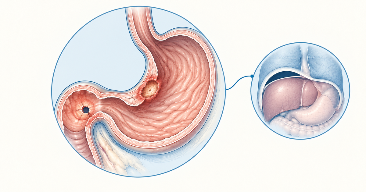
Free perforation usually involves the anterior duodenal wall. Look for gas under the diaphragm on an erect chest X-ray, but a normal film does not exclude it.
ACG 2024 prefers 14-day bismuth quadruple therapy as first-line for treatment-naive H. pylori. PPI, clarithromycin and amoxicillin triple therapy is the older regimen.
Think of Zollinger-Ellison syndrome when ulcers are multiple, postbulbar or refractory, or when diarrhoea is present.
Perforation needs rapid resuscitation, antibiotics and surgery. Boey factors help estimate risk, and a Graham patch is the classic omental repair.
Peptic Ulcer: Causes and Core Concepts
Two causes account for most ulcers that form in the stomach or the first part of the duodenum: H. pylori infection and NSAID use. Sources disagree about which one leads, so for exam purposes remember both and do not rank them (StatPearls, Gastric Ulcer).
H. pylori is found in more than 50% of duodenal ulcer patients and 30-50% of gastric ulcer patients, according to the MSD Manual Professional. Figures vary between sources, so learn the direction of the numbers (common in both, a little more so in duodenal ulcer) rather than one exact percentage.
Duodenal ulcer patients have higher mean basal and peak acid output than people without ulcers, although the ranges overlap considerably. Symptomatic duodenal ulcer usually appears between 25 and 40 years of age (PMC8171156).
Haemorrhage is the most common complication. Perforation and gastric outlet obstruction are the other classic complications, and each can be an indication for surgery. For more practice on this area, see the general surgery GI practice questions.
Duodenal vs Gastric Peptic Ulcer: Comparison Table
The table below puts the main distinguishing points side by side. It is built from the MSD Manual Professional and the other sources named in this article. Cross-check it against a standard surgery textbook before you rely on it for the exam.
| Feature | Duodenal ulcer | Gastric ulcer |
|---|---|---|
| Site | First few centimetres of the duodenum | Stomach |
| Pain pattern | Absent on waking, appears mid-morning, eased by food, returns 2-3 hours after meals | Less consistent, eating sometimes makes it worse |
| H. pylori association | More than 50% of patients | 30-50% of patients |
| Malignancy | Malignant duodenal ulcer is extremely rare | Must exclude cancer |
| Biopsy | Generally not warranted | Biopsy the ulcer edges |
| Typical perforation site | Anterior wall of the duodenum | Less common than duodenal |
| Bleeding vessel | Gastroduodenal artery (or superior pancreaticoduodenal artery) from an ulcer in the posterior wall of the bulb | - |
Sources: MSD Manual Professional, Peptic Ulcer Disease; duodenal bleeding artery from a 2024 PMC review, PMC10761636.
How to Use the Table in a Stem
Start with the pain-food relationship, a useful discriminator. Pain that eases after a meal, or night pain, points to a duodenal ulcer. Pain that worsens with eating, especially with weight loss or age above 60, should prompt you to exclude a gastric ulcer or malignancy.
Then look at the complication. A sudden severe abdominal pain with a rigid abdomen in a known ulcer patient points to perforation, and the anterior duodenal wall is the usual site. Back pain from a posterior duodenal ulcer suggests penetration into the pancreas. Bleeding from a posterior bulbar ulcer, by contrast, comes from erosion into the gastroduodenal artery or its neighbouring branches.
Why Gastric Ulcers Need a Biopsy
Gastric ulcers need edge biopsy because some are malignant. Suspicion rises with age above 60, weight loss and symptoms that do not respond to treatment (MSD Manual Professional). The same logic applies in surgery: for large perforated gastric ulcers, WSES suggests resection with frozen-section pathology when possible (Tarasconi et al., WSES 2020).
Modified Johnson Classification of Gastric Peptic Ulcer
The modified Johnson classification groups gastric ulcers by site and, for some types, acid status. The summary below follows a 2025 systematic review (World J Gastrointest Surg 2025, PMC12019041).
| Type | Location | Acid status |
|---|---|---|
| I | Body, lesser curve or incisura | No acid hypersecretion |
| II | Gastric body ulcer plus a duodenal ulcer | Acid oversecretion |
| III | Pyloric channel, within 3 cm of the pylorus | Acid oversecretion |
| IV | Proximal, near the gastro-oesophageal junction | Not stated in the source reviewed |
| V | Throughout the stomach | Associated with chronic NSAID use |
This classification is summarised from a secondary review, not a textbook. Confirm Types IV and V in your surgery textbook before the exam.
Memory aid (not from a textbook): Types II and III come with acid oversecretion, Type I does not, and Type V goes with NSAIDs.
H. pylori Treatment in Peptic Ulcer
The ACG 2024 guideline names 14-day bismuth quadruple therapy as the preferred first-line regimen for treatment-naive patients when antibiotic susceptibility is unknown (Chey et al., ACG 2024). Newer guidance has moved away from the older triple regimen.
ACG 2024 Bismuth Quadruple Therapy
The regimen has four components, each given for 14 days. The ACG evidence-based summary of the guideline lists (ACG, Schoenfeld, Sept 2024):
- A standard-dose PPI twice daily
- Bismuth (subcitrate or subsalicylate) four times daily
- Tetracycline 500 mg four times daily
- Metronidazole 500 mg three to four times daily
The same summary advises against substituting doxycycline for tetracycline. Alternatives in the guideline, for patients without penicillin allergy, are rifabutin triple therapy and a dual regimen of a PPI or potassium-competitive acid blocker with amoxicillin, each given for 14 days. Clarithromycin- or levofloxacin-containing regimens are reserved for cases where susceptibility is confirmed.
Older Triple Therapy
The older textbook regimen is a PPI with clarithromycin and amoxicillin. Guidelines now favour bismuth quadruple therapy, and other consensus guidance, including Maastricht VI/Florence, also favours bismuth-based regimens, typically for 14 days, where clarithromycin resistance is high or unknown (PMC12831489, 2026; PMC13247866, 2026). If an exam option lists both, check how the question is worded: a textbook-based question may expect triple therapy, while a guideline-based question points to bismuth quadruple therapy.
Testing for H. pylori
Test timing matters because some drugs cause false negatives. Stop PPIs for about 2 weeks and antibiotics for about 4 weeks before testing (PMC11175606, 2024).
| Test | Key point |
|---|---|
| Urea breath test | Gold-standard non-invasive test |
| Monoclonal stool antigen | Valid alternative to breath test |
| Rapid urease test | Affected by PPIs, H2 blockers and antibiotics; not for use after treatment |
| Serology | Cannot confirm eradication because antibodies persist |
Source: PMC11175606; NIDDK.
A test of cure is recommended after treatment, and it should be done at least 4 weeks after the antibiotics finish, with the PPI stopped at least 2 weeks beforehand (ACG 2024; NIDDK).
Special Peptic Ulcers: Zollinger-Ellison, Curling and Cushing
Three special ulcers are worth recognising from the clinical picture. Each is linked to a clear trigger.
Zollinger-Ellison Syndrome
Zollinger-Ellison syndrome (ZES) is caused by a gastrinoma, usually in the pancreas or duodenum, that drives gastrin hypersecretion. It produces severe, refractory ulcers, reflux and diarrhoea. About 20-30% of cases occur with MEN1 (PMC12730841, 2025).
Fasting serum gastrin is the first test, although PPIs, H. pylori and atrophic gastritis can also raise it. A gastric pH of 2 or below supports ZES. In the secretin test, a gastrin rise of 120 pg/mL or more is highly specific. Treatment includes lifelong PPI therapy.
Consider ZES when ulcers are multiple, postbulbar or refractory, or when diarrhoea is present (MSD Manual Professional). On imaging, Radiopaedia notes that multiple postbulbar ulcers with thickened gastric folds raise the suspicion.
Curling Ulcer
A Curling ulcer is a stress-induced duodenal ulcer in burn patients. It arises from inflammation and ischaemia and can perforate. It was described by Curling in 1842 (PMC12489155).
Cushing Ulcer
A Cushing ulcer is linked to raised intracranial pressure, as in brain tumours, head trauma and other intracranial conditions. It is classically attributed to raised intracranial pressure with vagal stimulation and increased acid secretion; the mechanism is debated (Asian J Neurosurg 2015; Kumaria et al., 2024). For a quick split, a Cushing ulcer goes with intracranial disease and a Curling ulcer with severe, extensive burns (PMC9622379). Do not treat the site as an absolute for either.
More broadly, stress ulcer risk factors include shock, coagulopathy, mechanical ventilation, glucocorticoids, anticoagulants, antiplatelets and NSAIDs (PMC13653456, 2026). Recognising a critically ill, burned or neurosurgical patient is usually enough to identify the type of ulcer in a stem.
Perforated Peptic Ulcer: Gas Under the Diaphragm
Free air under the diaphragm on an erect chest X-ray is the classic sign of a perforated peptic ulcer. Erect chest X-ray is probably the most sensitive plain radiograph for free intraperitoneal gas and can detect around 1 mL under the diaphragm (Radiopaedia, Pneumoperitoneum).
Free perforation usually occurs through the anterior duodenal wall, less often in the stomach (MSD Manual Professional). In a 703-patient perforation cohort, 56.5% were duodenal and 36.7% were gastric (Ann Gastroenterol Surg 2025, PMC12080193).
Erect Film Versus Left Lateral Decubitus
Patient position changes how little gas you can see. An upright chest film can show as little as about 1 mL of gas under the diaphragm (Radiopaedia). A cross-table left lateral decubitus film is an alternative, but it detects less gas than the erect film. Allow time for the gas to rise before you take the film.
A Normal Film Does Not Exclude Perforation
Free air is seen on X-ray in only 30-85% of perforations, according to WSES 2020, so a negative film does not rule it out (Tarasconi et al.). Free air together with a clear history and peritonitis justifies going to theatre. If there is no free air but suspicion persists, WSES suggests a water-soluble contrast study (weak recommendation).
WSES recommends CT for suspected perforation in an acute abdomen (Tarasconi et al.), but up to 12% of perforations may have a normal CT (Tarasconi et al.). A chest or abdominal X-ray is the initial test if CT is not promptly available.
Rigler Sign and Falciform Ligament Sign
The Rigler (double-wall) sign shows gas on both sides of the bowel wall on an abdominal radiograph. It appears only with a large volume of free gas (over about 1000 mL) and can be mimicked by touching bowel loops (Radiopaedia, Rigler sign). Do not rely on it to exclude perforation.
On a supine film, free gas that outlines the falciform ligament produces the falciform ligament sign, also known as the Silver sign. Because it requires a large pneumoperitoneum, it seldom occurs in isolation: a volume of gas that large usually also produces the Rigler sign (Radiopaedia, Falciform ligament sign).
Perforated Peptic Ulcer: Management and Surgical Repair
Management of a perforated ulcer rests on early resuscitation, antibiotics and operative repair. WSES 2020 suggests resuscitating rapidly, ideally within one hour, aiming for a mean arterial pressure of at least 65 mmHg, urine output of at least 0.5 ml/kg/h and normalising lactate (Tarasconi et al., WSES 2020). Broad-spectrum antibiotics are recommended, with a short course of 3-5 days suggested.
Delay costs lives. In a Danish cohort of 2,688 patients, each hour of surgical delay was associated with an adjusted 2.4% decrease in survival probability (Buck et al., cited in WSES 2020).
Non-Operative Care and Approach
WSES does not suggest routine non-operative management of perforation. Only extremely selected cases that seal on a contrast study qualify. For surgery, a laparoscopic repair is suggested for stable patients and open surgery for unstable patients.
Repair Choice and the Graham Patch
For perforations under 2 cm, WSES suggests primary repair. It makes no recommendation on whether an omental patch adds protection and does not recommend routine patching. For perforations of 2 cm or more, the plan depends on the site: resection with frozen section for large gastric ulcers, and resection or repair (with or without pyloric exclusion) for large duodenal ulcers.
The Graham patch is the classic textbook repair, and the Graham omental patch is described as the standard operation for perforated peptic ulcer (PMC12360380, 2025). It closes the perforation with an omental patch, and the Cellan-Jones repair is the pedicled omental alternative (PMC3952291, 2014). In the 2025 cohort above, Graham patch and Cellan-Jones repair were grouped as "plugging" and used in 244 of 538 operated patients (45.4%) (PMC12080193).
Omental patch leak rates of up to 12% have been reported for large ulcers, which is one reason large perforations call for a different plan (Tarasconi et al., WSES 2020).
Boey Risk Factors
The Boey score is a quick bedside estimate of risk in a perforated ulcer. It rests on three risk factors (PMC3952291, 2014):
- Concurrent severe medical illness
- Preoperative shock
- Longstanding perforation (more than 24 hours)
WSES suggests using Boey, PULP and ASA scores to assess risk (Tarasconi et al., WSES 2020). Boey is the most frequently used score, but its accuracy varies (PMC12360380, 2025). Hypoalbuminaemia is the strongest single predictor of mortality in the WSES guideline (Tarasconi et al., WSES 2020).
Memory aid (not from a textbook): Boey asks "Sick, shocked, or sat for a day?"
Putting Peptic Ulcer Notes Into Practice
Work through the topic in the order you would meet it in a patient: classify the ulcer, identify the cause, treat H. pylori correctly, and recognise perforation early. Then test yourself with timed questions, such as the gastroenterology practice set and the wider NEET PG question bank.
Conclusion
Peptic ulcer questions reward a clinical habit more than raw recall: read the stem for the trigger, the pain behaviour and the complication before you look at the options. Practise that reading on mixed question sets, and revisit any item where the guideline answer and the older textbook answer pointed in different directions.
Practise this on Reflex
Turn what you just read into recall with 14 years of tagged PYQs.
FAQ
Frequently asked questions
The questions aspirants ask most about this topic.
For a patient with no earlier H. pylori treatment and unknown antibiotic susceptibility, ACG 2024 recommends bismuth quadruple therapy for 14 days. The regimen is tetracycline 500 mg four times daily and metronidazole 500 mg three to four times daily, plus bismuth four times daily and a PPI taken twice daily. Clarithromycin-based triple therapy (PPI, clarithromycin and amoxicillin) is no longer the preferred first-line option; clarithromycin regimens are reserved for cases with confirmed susceptibility.
No. WSES 2020 reports free air on X-ray in 30-85% of perforations, so a negative film does not rule it out. WSES suggests a water-soluble contrast study when there is no free air but suspicion persists, and it recommends CT for suspected perforation in an acute abdomen. Up to 12% of perforations may have a normal CT.
Duodenal ulcer pain is absent on waking, appears mid-morning, is relieved by food and returns 2-3 hours after meals. Night pain is common. Gastric ulcer pain is less consistent, and eating sometimes makes it worse.
Gastric ulcers can be malignant, so the edges are biopsied, especially with age above 60, weight loss or symptoms that do not improve. Malignant duodenal ulcers are extremely rare, so duodenal biopsy is generally not warranted.
The Boey score is used to estimate risk in a perforated peptic ulcer. It looks at three things: severe medical illness alongside the perforation, shock before surgery, and a perforation that has gone on for more than 24 hours. WSES suggests the Boey, PULP and ASA scores for this purpose; Boey is the most widely used, though its accuracy varies.
Consider it when ulcers are multiple, postbulbar or refractory to treatment, or when diarrhoea is present. Fasting serum gastrin is the first test, and a gastric pH of 2 or below supports the diagnosis.
Stay ahead in your preparation
Get expert tips, exam updates, and high-yield insights delivered straight to your inbox.
No spam. Unsubscribe anytime.
More in Subject Notes
View all → Surgery
Burns for NEET PG: The Rule of Nines, Burn Depth and the Parkland Formula Worked Step by Step
 Surgery
Compartment Syndrome for NEET PG: Why Pulselessness Is the Sign That Comes Last, Not First
 Surgery
Hernia Types and Classification for NEET PG
Direct vs indirect inguinal hernia, Hesselbach’s triangle, and why femoral hernias strangulate most.
 Community Medicine (PSM)
Sensitivity and Specificity for NEET PG: A Worked 2x2 Table, PPV, NPV and the Prevalence Effect
Learn sensitivity and specificity through one worked 2x2 table, see why PPV and NPV move with prevalence, and sort out series versus parallel testing.