Placenta Previa vs Abruptio Placentae for NEET PG: The Painless vs Painful Bleeding Table
Reflex · 28 Oct 2026 · 14 min read
Last updated: 28 Oct 2026
On this page
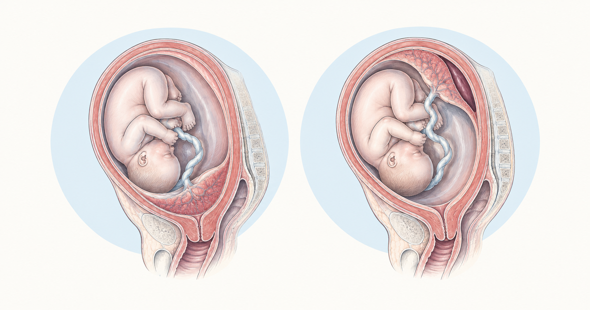
Placenta previa and abruptio placentae are the two classic causes of antepartum hemorrhage (bleeding in the second half of pregnancy) that students are asked to tell apart. They look alike on a quick read, because both present with bleeding late in pregnancy. The key distinction is one detail: is the bleeding painless, or is the uterus painful and tender?
This guide sorts the two conditions side by side. You will get definitions, risk factors, the one-line clinical difference, diagnosis rules, management and the key complications (Couvelaire uterus and DIC).
Key Takeaways
Think of previa when a woman bleeds suddenly and painlessly from the vagina in late pregnancy. The cause is a placenta that lies over some portion of the internal cervical os.
In abruptio placentae, a normally sited placenta detaches early. The uterus is painful, tender and irritable, and the blood may stay concealed.
When a woman bleeds late in pregnancy, avoid digital vaginal examination until previa has been ruled out. A speculum examination is safe.
Transvaginal ultrasound diagnoses previa. Abruption is diagnosed clinically, and a normal scan cannot rule it out.
A previous cesarean section raises the risk of both previa and accreta. For abruption, the key risks are hypertension, pre-eclampsia, prior abruption, trauma, smoking and cocaine.
Abruption can cause DIC, Couvelaire uterus and fetal death. Visible blood loss underestimates the real loss.
Placenta Previa: Definition and Types
The defining feature of placenta previa is a placenta that overlies the internal cervical os, either partly or completely (MSD Manual; Radiopaedia). Current practice classifies the placenta by its distance from the os rather than by older grades. Learn the threshold in both units, because an exam option may state it in either centimetres or millimetres ("within 2 cm" and "within 20 mm" mean the same thing).
Current classification
Radiopaedia describes three categories based on the lower placental edge:
| Category | Placental edge relative to internal os |
|---|---|
| Placenta previa | Covers any part of the internal os |
| Low-lying placenta | Edge less than 2 cm (20 mm) from the os but not covering it |
| Normal placenta | Edge more than 2 cm from the os |
Do not diagnose previa before 16 weeks. The lower segment grows during pregnancy, and an early low placenta often seems to "migrate" upward away from the os. As Radiopaedia notes, this apparent migration is attributed to trophotropism.
Older grading for recognition
Older textbooks list four types: Type I (low-lying), Type II (marginal), Type III (incomplete or partial central) and Type IV (complete or total central). Sources disagree on how these map to the Browne grades, and Radiopaedia says the four grades were hard to separate and were replaced. Learn the names so you can recognise them in an option list, but use the distance-from-os system for reasoning.
An even older split into "major" and "minor" previa is mentioned in Neilson's 2003 Cochrane review. Again, this is mostly historical.
How common is it?
Radiopaedia gives roughly 1 in 200 pregnancies, and the MSD Manual gives about 5 per 1,000 deliveries. A Cochrane review reports that around 5% of women have a low-lying placenta at 16 to 18 weeks, but only about 0.5% have previa at delivery. The MSD Manual adds that early previa usually resolves by 28 weeks.
Risk Factors for Placenta Previa
Most previa risk factors involve scarring or damage to the uterine lining, or a larger placental surface. The MSD Manual lists multiparity, prior cesarean delivery, uterine abnormalities (fibroids, prior curettage), prior uterine surgery such as myomectomy or multiple D&C, smoking, multiple gestation and older maternal age. Radiopaedia adds previous previa, assisted conception, a large placenta and previous manual removal of the placenta.
The risk factor to remember above all is a previous cesarean section. A registry study of 399,674 women (Gurol-Urganci et al., BMC Pregnancy and Childbirth, 2011) recorded previa in 8.7 per 1,000 second births when the first birth was a cesarean, against 4.4 per 1,000 when the first birth was vaginal. After adjustment, the odds ratio came to 1.60 (95% CI 1.44 to 1.76). The same paper pooled 37 studies and found an odds ratio of 2.20 (95% CI 1.96 to 2.46).
Placenta accreta becomes more likely with every further cesarean in a woman who has previa. Per the MSD Manual, accreta occurs in 3% of women with previa and one prior cesarean, 11% with two, 40% with three, 61% with four and 67% with five or more. For exams, the direction of the trend (more scars, more accreta) matters more than each percentage.
Placenta Previa Clinical Features: Painless Bleeding
The classic presentation is sudden, painless vaginal bleeding in the second or third trimester, which can be heavy enough to cause hemorrhagic shock (MSD Manual). Many women have no symptoms, and the previa is found on a second-trimester scan. Bleeding may start as early as 16 weeks, and Radiopaedia describes it as commonly occurring around 34 to 38 weeks.
The uterus is soft and non-tender on palpation, and the presenting part is often high because the placenta occupies the lower segment (Radiopaedia). Some women do have contractions (MSD Manual), so painless is the classic pattern, not an absolute rule. Fetal distress is usually late and follows maternal hemorrhage.
A simple memory aid (not a standard textbook mnemonic): "Previa = Pain-free, Abruption = Agony."
Abruptio Placentae: Definition, Risk Factors and Grades
Abruptio placentae is the premature separation of a normally implanted placenta, usually after 20 weeks and before the third stage of labour (MSD Manual; Radiopaedia). It can occasionally happen earlier. Incidence is roughly 0.4 to 1.5%, with a peak at 24 to 26 weeks according to the MSD Manual. Radiopaedia and GLOWM give about 1%.
Risk factors
The MSD Manual lists older age, chronic or pregnancy-related hypertension, placental ischemia (including IUGR), chorioamnionitis, vascular disease, prior abruption, abdominal trauma, thrombotic disorders, tobacco use, PPROM (especially with polyhydramnios) and cocaine use. Radiopaedia notes that pre-eclampsia or hypertension is present in up to 50% of cases.
A 2024 BJA Education review (Smith and Lee) puts numbers on the strongest risks. Previous abruption carries an odds ratio of 8 to 12, with a recurrence rate of 4.4% after one abruption and 19 to 25% after two or more. Cocaine or amphetamine use carries an odds ratio of 5 to 10. The same review notes that about 70% of cases occur in women with no obvious risk factor, so a low-risk history does not rule abruption out.
Clinical features
Abruption differs from previa in the uterus. It is painful, tender and irritable, and the pain can be out of proportion to the visible bleeding (MSD Manual). Bleeding may be absent when the blood is concealed behind the placenta. In severe cases, shock, DIC and fetal distress or death can follow. Chronic abruption can instead show intermittent dark brown spotting.
About 80% of abruptions have external bleeding and about 20% are concealed, according to the GLOWM chapter. Because some of the blood stays concealed, visible loss underestimates the total loss.
Grading (Sher and the textbook system)
The BJA Education review describes Sher staging. Stage I is mild, with a small retroplacental hematoma often found only after delivery. Stage II is intermediate, with a hypertonic uterus and a live fetus. Stage III is severe, with intrauterine fetal death, split into IIIa (no coagulopathy) and IIIb (coagulopathy).
The GLOWM chapter gives a three-grade system with similar logic:
| Grade | Share of cases | Key features |
|---|---|---|
| I | About 40% | No coagulopathy or fetal distress |
| II | About 45% | Hypertonic uterus, hypofibrinogenemia, fetal distress, live fetus |
| III | About 15% | Dead fetus, hypotension, thrombocytopenia, tetanic uterus; IIIA no overt coagulopathy, IIIB overt coagulopathy |
You may also meet the Page classification (grades 0 to 3) in exam-prep material. Standard references such as the MSD Manual and Radiopaedia, and the reviews used here, do not name it, so check your standard textbook for the exact definition before relying on it.
Placenta Previa vs Abruptio Placentae: The Comparison Table
The table below is the high-yield summary. Reproducing it from memory is a quick way to test your grasp of the differences.
| Feature | Placenta previa | Abruptio placentae |
|---|---|---|
| Bleeding | Sudden, painless | Painful; may be concealed |
| Uterus | Soft, non-tender; presenting part high | Tender, irritable, hypertonic |
| Fetal distress | Usually late, from maternal hemorrhage | Common; fetal death in grade III |
| Blood loss | Visible bleeding | Visible loss underestimates total loss |
| Key risks | Prior cesarean, multiparity, smoking, multiple gestation, older age | Hypertension or pre-eclampsia, prior abruption, trauma, smoking, cocaine |
| Diagnosis | Transvaginal ultrasound; no digital examination | Clinical; ultrasound is insensitive |
| Delivery | Cesarean; 36 to 37 6/7 weeks if stable | Cesarean if unstable or term; vaginal delivery possible if stable |
Sources for the table: MSD Manual (previa and abruption chapters), Radiopaedia, GLOWM and the 2024 BJA Education review.
Worked scenario
What would you do for a G2 woman at 32 weeks, with one earlier cesarean section, who arrives after a sudden bleed that caused her no pain, and whose uterus is soft and non-tender on palpation? Painless bleeding together with a prior cesarean, a key risk factor, fits placenta previa in the table above. Do not perform a digital vaginal examination until previa has been excluded. The best next step is a transvaginal ultrasound to locate the placenta, and a speculum examination can be done safely. This is a practice scenario built from the sourced points above, not a recalled exam question.
To test the same reasoning on more cases, try the OBG obstetric complications practice questions.
Diagnosing Placenta Previa and Abruption
Previa is diagnosed by imaging, whereas abruption is diagnosed by examination and context. The first safety rule is the same for both: do not perform a digital vaginal examination after 20 weeks unless previa has been excluded (MSD Manual). A speculum examination is safe.
Placenta previa
Transvaginal ultrasound (TVS) is accurate and safe, and it is superior to transabdominal and transperineal scanning, as summarised from the RCOG guideline by Medical Dialogues. Transabdominal scanning can overdiagnose previa. A full bladder or a myometrial contraction can mimic previa, so take postvoid images. A subchorionic hematoma over the cervix can also mimic it. Radiopaedia calls MRI the gold standard, though it is seldom required.
For follow-up, the RCOG guideline as summarised by Medical Dialogues advises a repeat TVS at 32 weeks if a low-lying placenta or previa is seen at the anomaly scan, and again at about 36 weeks if it is still low and the woman is asymptomatic.
Abruptio placentae
Abruption is a clinical diagnosis (MSD Manual). Ultrasound has a specificity of 96% but a sensitivity of only 24% (BJA Education). A retroplacental hematoma is visible in only 2 to 25% of cases (Radiopaedia). A normal scan therefore never excludes abruption.
The workup in the MSD Manual includes fetal heart rate monitoring, CBC, blood group and Rh typing, PT and PTT, fibrinogen and fibrin degradation products. A Kleihauer-Betke test is done in Rh-negative women to size the anti-D dose. Rule out previa before any pelvic examination.
Managing Placenta Previa
Management depends on how much the woman is bleeding and how far along the pregnancy is. The MSD Manual recommends immediate cesarean delivery for heavy or uncontrolled bleeding, a nonreassuring fetal heart rate or maternal instability.
Stable women
After a first bleed before 36 weeks, the MSD Manual advises hospitalisation, modified activity, no sexual activity and Rho(D) immune globulin if the woman is Rh-negative. While she stays stable, the plan is a cesarean at 36 to 37 6/7 weeks. The RCOG guideline, as summarised by Medical Dialogues, suggests considering 36+0 to 37+0 weeks for uncomplicated previa and 34+0 to 36+6 weeks if there is bleeding or another preterm risk. These windows overlap without matching exactly, so quote the one given in your own textbook.
Steroids and tocolysis
The RCOG summary gives a single course of antenatal corticosteroids at 34+0 to 35+6 weeks, earlier if preterm delivery risk is higher. The MSD Manual says some experts give steroids if delivery may be needed before 34 weeks, and consider them at 34 to 36 weeks if not given earlier. Tocolysis can be used for up to 48 hours in symptomatic women to allow steroids to act. When delivery is indicated, tocolysis must not be used to delay it.
Mode of delivery
Cesarean delivery is the rule for previa. The MSD Manual allows vaginal delivery only for a low-lying placenta with its edge within 1.5 to 2.0 cm of the os, after shared decision-making.
Expectant management and its history
The Cochrane review by Neilson (2003) found insufficient trial evidence to change practice for expectant management. It also notes that Macafee began conservative management in Belfast in 1945, when maternal mortality from previa was as high as 5%. In exam terms, expectant (Macafee) management means in-hospital observation until fetal maturity. It is a historical concept, and current practice follows the timing above.
Accreta spectrum
Women with a prior cesarean and an anterior low-lying placenta or previa should be screened for accreta. The RCOG summary advises delivery in a specialist centre, and planned delivery at 35+0 to 36+6 weeks. If accreta is not suspected until after birth, the placenta is left in situ.
Managing Abruptio Placentae
Abruption management follows maternal and fetal stability. The MSD Manual recommends cesarean delivery for maternal instability, a nonreassuring fetal heart rate or term gestation (37 weeks or more). Vaginal delivery is reasonable if the mother is stable and the fetal heart rate is reassuring.
For preterm abruption (under 37 weeks) that is not life-threatening, with a reassuring fetal heart rate, the woman is hospitalised and observed. Steroids are given below 34 weeks and selectively at 34 to 36 weeks. In chronic abruption with reassuring status, the BJA Education review suggests expectant management with induction or cesarean at about 37 weeks.
In shock, the first step is rapid crystalloid infusion with close monitoring. Remember that concealed loss is underestimated (GLOWM). For major hemorrhage, the BJA Education review describes tranexamic acid 1 g IV over 10 minutes when blood loss exceeds 1,000 mL, and fibrinogen replacement if fibrinogen is 2 g/L or lower.
Couvelaire Uterus and DIC in Abruption
Couvelaire uterus (uteroplacental apoplexy) is blood spreading through the myometrium and serosa, and it can dissect into the broad ligaments (GLOWM). The serosa and broad ligaments take a dark violaceous hue. It is listed as a complication of abruption by Radiopaedia.
DIC follows when thromboplastin enters the circulation and activates clotting. Fibrinogen is consumed, and secondary fibrinolysis produces fibrin degradation products (GLOWM; BJA Education). DIC occurs in about 10% of abruptions, mostly severe ones (GLOWM).
The tests differ in sensitivity. PT and PTT are insensitive, serial fibrinogen is sensitive, and FDPs or D-dimer may be the most sensitive (GLOWM). In pregnancy, a fibrinogen below 200 mg/dL is abnormal (GLOWM). The BJA Education review gives a normal range of 4 to 6 g/L and notes that below 2 g/L predicts severe hemorrhage.
Abruption can also cause fetal growth restriction and fetal demise. The BJA Education review reports fetal mortality of 3 to 12% with abruption, against 0.6% without it. Previa complications are different: antepartum hemorrhage, a high presenting part, accreta spectrum and maternal or fetal compromise (Radiopaedia).
Exam Pitfalls for Placenta Previa and Abruption
Most wrong answers come from a small set of traps. Check yourself against this list:
- Never do a digital examination before previa is excluded, and rule out previa before any pelvic examination in suspected abruption.
- A normal ultrasound does not exclude abruption.
- Visible blood underestimates loss in abruption.
- Do not diagnose previa before 16 weeks, because of apparent placental migration.
- "Within 2 cm" and "within 20 mm" are the same threshold for a low-lying placenta.
- Rh-negative women need anti-D in both conditions, and a Kleihauer-Betke test helps size the dose in abruption.
- Painless bleeding is classic for previa but not absolute, since some women with previa have contractions.
Conclusion
Placenta previa and abruptio placentae both cause bleeding in late pregnancy, but they separate cleanly on pain, uterine tone, risk factors and diagnosis. Previa is painless with a soft uterus and is confirmed by transvaginal scan. Abruption is painful with a tender, hypertonic uterus, is diagnosed clinically and can bring DIC and Couvelaire uterus. Practise the comparison table until you can write it unaided. For related OBG topics, see our notes on types of abortion and the partograph.
Practise this on Reflex
Turn what you just read into recall with 14 years of tagged PYQs.
FAQ
Frequently asked questions
The questions aspirants ask most about this topic.
Painless, sudden vaginal bleeding with a soft, non-tender uterus is the classic picture of placenta previa. In abruptio placentae the uterus is painful, tender and irritable, and the blood may be concealed. Abruption pain can be out of proportion to the bleeding you can see.
A digital examination can disturb a placenta lying over the cervix and provoke heavy bleeding. It should not be done after 20 weeks unless placenta previa has been excluded. A speculum examination is safe and can be used to look at the cervix.
Transvaginal ultrasound is the preferred test because it is accurate and safe. Transabdominal scanning can overdiagnose previa, and a full bladder or uterine contraction can mimic it, so postvoid images help. Do not diagnose previa before 16 weeks, since the placenta can seem to move away from the os as the uterus grows.
No. Abruption is a clinical diagnosis. One 2024 review reports ultrasound specificity of 96% but sensitivity of only 24%, and a retroplacental hematoma is seen in only a minority of cases. A normal scan therefore never excludes abruption.
Couvelaire uterus, also called uteroplacental apoplexy, is a complication of abruption in which blood spreads through the myometrium and serosa and may dissect into the broad ligaments. The uterine serosa and broad ligaments take on a dark violaceous colour.
The MSD Manual recommends cesarean delivery at 36 to 37 6/7 weeks for stable women. The RCOG guideline, as summarised by Medical Dialogues, suggests 36+0 to 37+0 weeks for uncomplicated previa and an earlier window of 34+0 to 36+6 weeks if there is bleeding or another preterm risk. Immediate cesarean is needed for heavy bleeding, a nonreassuring fetal heart rate or maternal instability.
Stay ahead in your preparation
Get expert tips, exam updates, and high-yield insights delivered straight to your inbox.
No spam. Unsubscribe anytime.
More in Subject Notes
View all → Obstetrics & Gynaecology
Partograph for NEET PG: What the Alert Line and Action Line Actually Mean
 Obstetrics & Gynaecology
Types of Abortion for NEET PG: Sorted by Cervix Status and What's Actually Been Passed
 Obstetrics & Gynaecology
APGAR Score for NEET PG: Components, Scoring, and What It Predicts
All five components, the exact scoring criteria, and why the 5-minute score matters more than the 1-minute.
 Community Medicine (PSM)
Sensitivity and Specificity for NEET PG: A Worked 2x2 Table, PPV, NPV and the Prevalence Effect
Learn sensitivity and specificity through one worked 2x2 table, see why PPV and NPV move with prevalence, and sort out series versus parallel testing.