Types of Wounds for NEET PG: Mechanical Injuries, Medicolegal Points and BNS Grievous Hurt
Reflex · 4 Nov 2026 · 14 min read
Last updated: 4 Nov 2026
On this page
A wound is a break in the continuity of tissue caused by violence. In forensic medicine the useful question is never just "what is this wound called?" but "what weapon made it, how did it happen, and what does it mean in law?" This guide sorts mechanical injuries by the force that caused them, then links each type to its medicolegal use.
Here each type is tied to the three questions a medicolegal report must answer: the weapon, the manner (accident, suicide or homicide) and the legal category of hurt.
Key Takeaways
Mechanical wounds fall into two broad groups: blunt force (abrasion, contusion, laceration) and sharp force (incised, stab, chop).
Margins and bridges separate a tear from a cut: a lacerated wound has ragged, crushed edges crossed by tissue bridges, while an incised wound has clean-cut edges, no bridges and a surface length greater than its depth. A stab wound reverses that proportion, with depth exceeding surface length.
A bruise cannot be aged reliably from its colour. The best evidence is paediatric and dates from 2005, so treat textbook colour sequences as a rough guide only.
Hesitation marks point towards self-infliction, and defence wounds point towards an assault, but neither is conclusive alone.
Under the Bharatiya Nyaya Sanhita (BNS) 2023, section 114 defines hurt and section 116 lists eight kinds of grievous hurt, one of which turns on fifteen days of severe pain or inability to follow ordinary pursuits.
How are the types of wounds classified?
The simplest working classification is by the type of force. Blunt force produces abrasions, contusions and lacerations. Sharp force produces incised wounds, stab wounds and chop wounds. Firearm injuries are usually studied as a separate group because they combine features of both. StatPearls describes the basic blunt-force trio in these terms, as summarised in its blunt force trauma chapter.
A quick way to hold the scheme in your head:
- Blunt: abrasion (surface scrape), contusion (bleeding under intact skin), laceration (tear).
- Sharp: incised (cut), stab (puncture), chop (heavy cutting weapon).
- Firearm: entrance and exit wounds, classified by range.
The same wound can carry several labels. A single blow with a blunt object can produce an abrasion at the surface, a contusion beneath it and a laceration where the skin splits. Describe each component separately in a report.
Abrasion wounds: the surface scrape
An abrasion is the loss of the superficial epidermis through scraping, impact or pressure. It heals without scarring unless it reaches the dermis, and StatPearls notes that healing takes about two weeks (abrasion chapter). The same source stresses that the nature of an abrasion matters more medicolegally than its severity.
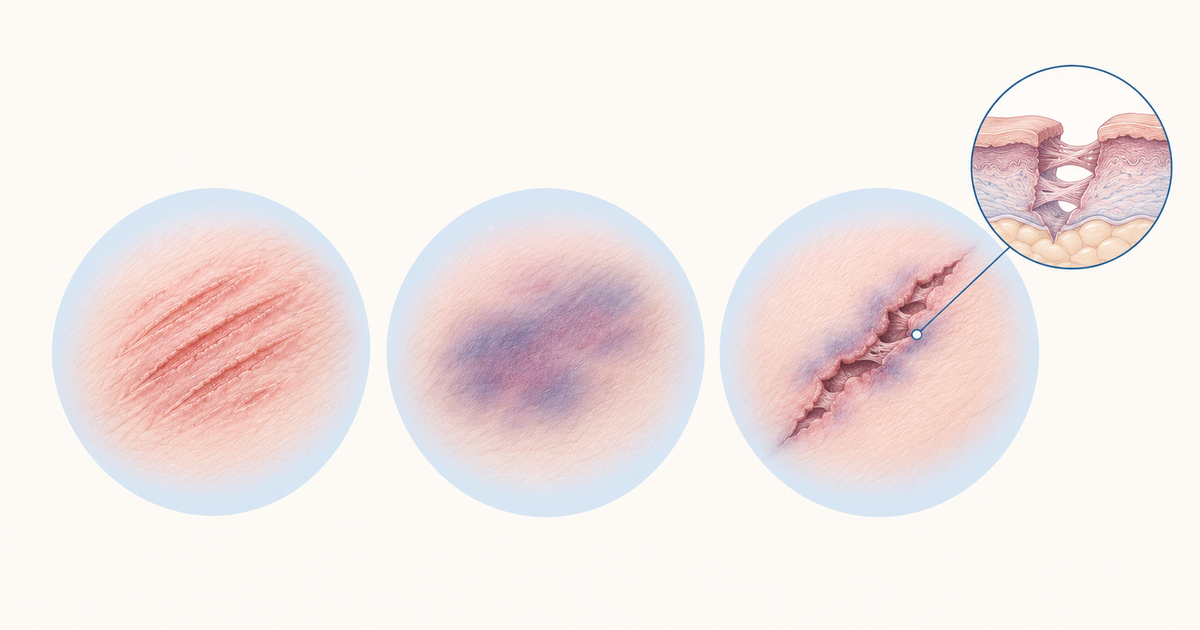
That is the key idea. A small abrasion can carry a pattern: the shape of a ligature, a fingernail or a tyre can be imprinted on the skin. Textbooks group abrasions as scratches, grazes, pressure abrasions and impact abrasions. Learn the group by the mechanism that produces it: a sharp point dragged across skin, a sliding contact, a sustained pressure and a direct blow.
Abrasions matter because they are often the only visible sign of the force applied. In a strangulation or a road traffic injury, the surface pattern may be the best clue to what touched the body.
Contusion wounds: bleeding under intact skin
A contusion (bruise) is the rupture of small vessels beneath intact skin, with blood leaking into the tissues. The skin surface is not broken, which separates it from an abrasion or a laceration. Textbooks distinguish a bruise from a haematoma, where the blood collects as a larger pooled mass, and from ecchymosis, which is a diffuse area of bleeding.
Blood in a deep bruise can track along tissue planes, so the visible mark may appear away from the point of impact. This is why a mark on the skin does not always tell you exactly where the blow landed.
Can you age a bruise from its colour?
No, not reliably. This is a point where common teaching runs ahead of the evidence. Maguire and colleagues reviewed 167 studies on ageing bruises in children and included only three. They found that colour alone was unreliable, and that every colour appeared in bruises of every age.
The accuracy figures are poor. Clinicians placed a bruise within 24 hours of its true age correctly less than 40% of the time, and classed bruises as fresh, intermediate or old correctly only 55 to 63% of the time. The review concluded that a bruise cannot be accurately aged from clinical assessment in vivo or from a photograph (Maguire et al., Arch Dis Child 2005).
Two limits apply to this evidence. It covers children only, and its searches ran to 2004. Even so, it is the best sourced statement available here, and it is enough to say that the textbook sequence (red or purple early, yellow or green later) is a rough guide, not a clock.
Some bruise-ageing details in the same review are narrow: yellow was first seen at about 24 hours in one study and within 48 hours in others, and red was seen only in bruises of roughly a week or less. Treat those as study findings, not rules.
In practice, write what you see, avoid giving an exact age and say that colour is an unreliable marker.
Lacerated wounds: the tear
A laceration is a tear of tissue caused by blunt force that crushes or stretches the skin until it splits. Its hallmark is tissue strands (bridges) that cross the wound, and the margins are crushed and ragged. StatPearls describes it as a tear with tissue strands bridging the wound.
Textbooks describe subtypes including split lacerations (skin crushed against bone, typical over the scalp or shin), avulsions (skin torn off in a flap or sheet), cut lacerations and tears. Learn the subtypes by the mechanism: crushing against bone, shearing, a sharp-edged blunt object and overstretching.
Why a split laceration can look like a cut
A split laceration over bone, such as the scalp, can have fairly straight edges and look like an incised wound. The distinguishing features are the crushed margin and the tissue bridges. A case report describes an atypical lacerated abdominal wound that mimicked an incised wound, so bridging should be read together with the other features, not as an absolute sign (PubMed 23424938).
Types of wounds: incised, stab and chop
Sharp-force wounds are produced by an edge or a point. The three types are separated by their dimensions and the weapon. Use the table as the core comparison.
| Feature | Incised wound | Lacerated wound | Stab wound |
|---|---|---|---|
| Force | Sharp edge drawn across skin | Blunt force crushing or stretching | Pointed weapon driven in |
| Dimension | Length exceeds depth | Variable | Depth exceeds length |
| Margins | Clean cut, intact epidermis to the edge | Ragged, crushed, abraded | Usually clean, shape reflects the blade |
| Tissue bridges | Absent | Present | Absent |
| Hair bulbs | Cleanly cut | Crushed | Not a distinguishing feature |
| Bleeding | Free | Often less than expected | Depends on the vessels reached; deep injury may be mostly internal |
The lacerated column and the incised column follow the operating-microscope study that contrasted the two: lacerations show a crushed epidermis at the margin and bridging tissue or vessels, while incised wounds have intact epidermis to the edge and no bridges (PubMed 15335652). The remaining row details come from standard textbook teaching.
Incised wounds
Clean-cut edges, no bridging and cleanly cut hair bulbs mark an incised wound, whose surface length exceeds its depth. Bleeding tends to be free, because cleanly divided vessels do not retract and seal as readily as torn ones. In self-harm, the neck and wrist are the classic sites for such a cut.
Stab wounds
A pointed weapon makes a stab wound, whose depth exceeds its length on the skin. The description depends on the number of cut edges the blade has. Two terms are worth keeping apart: a penetrating wound has an entry only, and a perforating wound has both an entry and an exit.
The external size of a stab wound can mislead. The depth of the track and the structures reached decide the danger, so the skin wound may look small while the internal injury is severe.
Chop wounds
A chop wound is made by a heavy sharp-edged weapon such as an axe. It cuts deep into the tissues and often damages the underlying bone. Think of it as a hybrid: the edge is sharp, but the weight behind it adds a crushing component.
Hesitation marks and defence wounds
Hesitation marks and defence wounds are the two patterns that help reconstruct the manner of an injury. A comparison study of 58 suicides and 149 homicides found hesitation marks in 43 suicides (about 74%) and defence wounds in 91 homicides (about 61%) (PubMed 19291442). Those figures show that neither finding is universal in its own group.
| Feature | Hesitation marks | Defence wounds |
|---|---|---|
| Meaning | Tentative, shallow cuts made before a deeper one | Injuries from the victim trying to ward off an attack |
| Suggests | Self-infliction | Assault by another person |
| Typical sites | Neck, left chest, flexor surface of the wrist or radial forearm | Hands, forearms and fingers |
| Distribution | Hand marks are usually one-sided | More often involve both hands |
| Character | Several tentative cuts, typically shallower than the fatal wound | Often incised wounds in knife attacks |
Textbooks split defence wounds into active (the victim grasps the blade) and passive (the victim raises the arm to shield the body). In a cross-sectional study of defence injuries in homicide, they were most often on the hands, forearms and fingers, and incised wounds dominated in knife attacks (PMC11964116).
Do not over-read the absence of a feature. A 2024 report describes a suicide by a single incising cut with no hesitation marks, a reminder that their absence does not exclude suicide (PubMed 38851015). The manner of death needs the scene, the history and toxicology considered together.
Firearm wounds: entrance, exit and range
Firearm wounds are classified by whether the projectile entered or left the body, and by how far the muzzle was from the skin. A 2026 narrative review of gunshot wound patterns summarises the features (Cureus 2026).
An entrance wound is generally smaller than the exit, with inverted edges. It often has an abrasion collar and may have an ecchymotic ring. An abrasion collar can also occur at an exit wound if the projectile meets resistance, such as tight clothing. Entrances from rifled firearms are often circular, especially contact shots not over bone.
An exit wound is larger, with everted and irregular margins. It lacks flame, singeing, tattooing or a grease collar, and it may contain clothing fibres or bone fragments.
The range categories rest on what the muzzle leaves on the skin:
| Range | What you see |
|---|---|
| Contact | Soot deep in the wound; a muzzle imprint is possible |
| Close | Soot and stippling; stippling is associated with firing within about 2 feet (60 cm) |
| Intermediate | Stippling or tattooing without soot |
| Distant | Neither soot nor powder reaches the body |
Cut-off distances depend on the weapon and ammunition, so the figures above are approximate. The same review notes that shotgun tattooing disappears beyond about 1 metre, and that an absence of residue alone does not prove a distant shot.
Wound vitality: was the injury made in life?
Vitality is a question of timing relative to death: was the person alive or dead when the injury was inflicted? Wound age is a separate question, the interval the person survived after being injured. A systematic review in the International Journal of Molecular Sciences makes the central point: histology alone is not enough to prove the vitality of a lesion, because the method is operator-dependent and prone to artefact. Early post-mortem processes and decomposition can mimic vital reactions (Int J Mol Sci 2022).
Immunohistochemistry is the most studied approach and is often called the gold standard, but it lacks standard protocols. Biochemical and molecular markers are not yet forensically reliable. Textbooks list ante-mortem signs such as haemorrhage, swelling, an inflammatory reaction and healing. Use them together, and do not rest a conclusion on one sign.
For the related post-mortem changes that run alongside wound interpretation, see postmortem lividity.
Types of wounds in law: hurt and grievous hurt under the BNS 2023
The medicolegal grading of an injury now follows the Bharatiya Nyaya Sanhita, 2023. Under section 114, anyone who causes bodily pain, disease or infirmity to another person is said to cause hurt. Section 116 defines grievous hurt, and section 117 deals with voluntarily causing it (BNS 2023 text, NCRB Sankalan Portal).
Section 116 contains eight clauses, (a) to (h). Grouped by theme, they are:
- Senses (clauses b and c): permanent privation of the sight of either eye, and permanent privation of the hearing of either ear.
- Members and joints (clauses d and e): privation of any member or joint, and destruction or permanent impairing of the powers of any member or joint.
- Other losses (clauses a and f): emasculation, and permanent disfiguration of the head or face.
- Bone and tooth (clause g): fracture or dislocation of a bone or tooth.
- Danger or prolonged disability (clause h): any hurt that endangers life, or that causes the sufferer to be, for fifteen days, in severe bodily pain or unable to follow his ordinary pursuits.
Learn the list as eight clauses in a fixed order: the first six are functional or structural losses, the seventh is a fracture or dislocation (see types of fractures for the bone-injury patterns), and the eighth is the catch-all based on danger to life or fifteen days of disability.
Memory aid (our own, not part of the statute): take the first word of each clause in order, Emasculation, Eye, Ear, Part lost, Power of part impaired, Disfigured head or face, Fracture, Hurt endangering life. A sentence that follows the same initials is "Eight Eager Elephants Pulled Pink Daisies From Hedges". Use it only to recall the order, then check each clause against the list above.
Older teaching used a three-way grading of slight, dangerous and mortal, but the current legal framework to use is the BNS sections above.
For a doctor, the practical point is that the wound's type does not decide its legal category. A stab wound may be simple hurt or grievous hurt, depending on what it did. The report should describe the injury and its effects, and the court decides the legal label.
Common mistakes when revising types of wounds
Several slips come from treating the labels as the goal.
- Reading colour as age. A yellow bruise is not automatically a day or two old, as shown above.
- Calling every straight-edged wound incised. Check the margins and bridges before deciding between incised and a split laceration.
- Judging a stab wound by its skin length. The track depth decides the danger.
- Treating hesitation marks as proof. They support self-infliction but do not prove it, and their absence does not exclude it.
- Using old section numbers. Use BNS 114, 116 and 117.
A useful drill is to draw the incised and lacerated wound edges from memory, label the bridges and the clean-cut hair bulbs, and then say aloud what each tells you about the weapon. Then work through scenario-based questions on the forensic medicine injuries practice page.
Types of wounds: what to carry into revision
Wounds are classified by force, but they are read by what they reveal: the weapon, the manner and the legal category of hurt. Keep the blunt and sharp groups apart, use margins and bridges to separate lacerated from incised, and remember that bruise colour and single signs such as hesitation marks are supporting clues, not proof. For the legal grading, anchor on BNS 114, 116 and 117. When you are ready to test yourself, move on to the NEET PG question bank.
Practise this on Reflex
Turn what you just read into recall with 14 years of tagged PYQs.
FAQ
Frequently asked questions
The questions aspirants ask most about this topic.
Mechanical wounds are grouped by force. Blunt-force wounds are abrasions, contusions and lacerations. Sharp-force wounds are incised, stab and chop wounds. Firearm wounds are usually studied as a separate group, with entrance and exit wounds classified by range.
An incised wound has clean-cut edges, intact epidermis to the margin, no tissue bridges and cleanly cut hair bulbs, and it is longer than it is deep. A lacerated wound has ragged, crushed margins with tissue bridges. Bridging alone is not absolute, because an atypical laceration can mimic an incised wound.
Not reliably. A 2005 review of paediatric studies found that colour alone was unreliable and that every colour appeared in bruises of every age. Textbook colour sequences are only a rough guide.
Hesitation marks are tentative, shallow cuts that suggest self-infliction, typically on the neck, left chest or wrist. Defence wounds are injuries from a victim warding off an attack, typically on the hands, forearms and fingers. Neither is conclusive alone.
Section 116 of the Bharatiya Nyaya Sanhita, 2023 defines grievous hurt through eight clauses. Clause (a) covers emasculation. Clauses (b) and (c) cover permanent privation of the sight of either eye or the hearing of either ear. Clauses (d) and (e) cover privation of any member or joint, and destruction or permanent impairing of the powers of any member or joint. Clause (f) covers permanent disfiguration of the head or face, and clause (g) covers fracture or dislocation of a bone or tooth. Clause (h) covers any hurt that endangers life, or that leaves the sufferer in severe bodily pain or unable to follow ordinary pursuits for fifteen days.
Stay ahead in your preparation
Get expert tips, exam updates, and high-yield insights delivered straight to your inbox.
No spam. Unsubscribe anytime.
More in Subject Notes
View all → Forensic Medicine
Rigor Mortis for NEET PG: The Timeline Every Time-of-Death Question Is Really Testing
 Forensic Medicine
Postmortem Lividity for NEET PG: Timeline, Color Clues, and Time of Death
Onset, fixation, the colour clues that flag poisoning, and how lividity helps estimate time since death.
 Anesthesia
ASA Physical Status Classification for NEET PG: Classes I to VI, Examples and the E Modifier
A clear walk through the ASA physical status classification: what each class means, example patients, the E modifier for emergencies, and the sorting mistakes to avoid.
 Psychiatry
Defense Mechanisms for NEET PG: Mature, Immature and Neurotic Defenses with One Example Each
Learn defense mechanisms as a ladder from pathological to mature, with one worked example per level, clear notes on projection and sublimation, and where Vaillant's scheme and the DMRS disagree.