Reed-Sternberg Cells and Hodgkin vs Non-Hodgkin Lymphoma: The Full NEET PG Comparison
By Dr. Utsav Bhattacherjee, MBBS, MBA · 2 Sept 2026 · 11 min read
Last updated: 23 Sept 2026
On this page
Hodgkin and non-Hodgkin lymphoma get tested as a pair almost as often as they get tested individually, and the fastest way to answer either question is to anchor on one single finding: the Reed-Sternberg cell. Its presence or absence is what actually separates the two categories — everything else in this comparison follows from that one fact.
Reed-Sternberg Cells: The Defining Feature
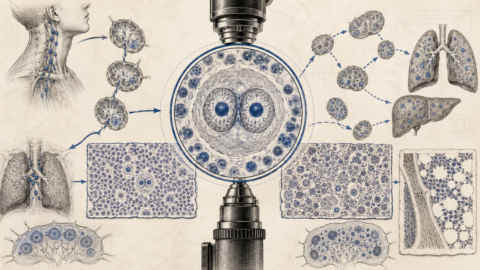
The Reed-Sternberg cell is a large, abnormal B-cell with a characteristic binucleate, "owl-eye" appearance — two mirror-image nuclei, each containing a large, prominent nucleolus, giving the cell its distinctive look under the microscope. Immunohistochemically, classic Reed-Sternberg cells are CD15-positive and CD30-positive, which is the marker combination examiners use to confirm the diagnosis when a vignette describes immunohistochemistry findings rather than just morphology.
A detail worth holding onto: Reed-Sternberg cells are typically a minority of the total cell population in the affected lymph node. The bulk of the tissue is made up of a reactive background — lymphocytes, eosinophils, plasma cells, and histiocytes recruited by cytokines the Reed-Sternberg cells themselves secrete. This is part of why Hodgkin lymphoma can look deceptively "inflammatory" on a low-power view, and why finding the Reed-Sternberg cells specifically, rather than just characterizing the overall picture, is the actual diagnostic step.
How the Diagnosis Is Actually Made
Diagnosis requires an excisional lymph node biopsy, not a fine-needle aspiration. This is a genuinely testable point on its own: FNA collects a suspension of individual cells, which is enough to sample many malignancies but not enough to classify a lymphoma correctly, since subtyping Hodgkin lymphoma depends heavily on the architecture of the node — whether the pattern is nodular or diffuse, how the background cell population is distributed, whether sclerotic bands are present. An excisional biopsy preserves that architecture; an FNA destroys it by design.
CD30 specifically has clinical relevance beyond confirming the diagnosis: brentuximab vedotin, an antibody-drug conjugate used in Hodgkin lymphoma treatment, is designed to specifically target CD30-positive cells. A question connecting the diagnostic marker to why that specific marker was chosen as a drug target is testing an understanding of the mechanism, not just recall of the immunophenotype.
The Subtypes of Classic Hodgkin Lymphoma
Nodular sclerosis is the most common subtype overall, and it has a distinctive demographic skew toward young adults, especially women. Histologically, it's defined by broad bands of collagen (sclerosis) dividing the node into nodules, and by a variant Reed-Sternberg cell called the lacunar cell — named for the artifactual clear space that forms around it during tissue processing. A mediastinal mass in a young woman is the classic clinical setup this subtype is written into.
Mixed cellularity shows a genuinely heterogeneous mix of inflammatory cells alongside more numerous classic Reed-Sternberg cells, and it skews toward an older population and toward HIV-positive patients specifically — a detail that shows up often enough in vignettes to be worth remembering as a distinguishing clue.
Lymphocyte-rich carries the best prognosis of the classic subtypes, dominated by reactive lymphocytes with relatively few Reed-Sternberg cells. Lymphocyte-depleted sits at the opposite end — the worst prognosis, relatively rare, with numerous Reed-Sternberg cells and few background lymphocytes, typically presenting in older or immunocompromised patients.
A useful shortcut for the two prognosis extremes: more lymphocytes relative to Reed-Sternberg cells generally tracks with a better prognosis, and fewer lymphocytes relative to Reed-Sternberg cells tracks with a worse one — the immune system's ability to mount a reactive response against the tumor cells is, roughly, what the lymphocyte population represents.
Nodular Lymphocyte-Predominant: The Separate Entity
Nodular lymphocyte-predominant Hodgkin lymphoma (NLPHL) is worth treating as genuinely distinct from the four classic subtypes above, not as a fifth variant of the same thing. Instead of classic Reed-Sternberg cells, it features "popcorn cells" (also called L&H cells, for lymphocytic and histiocytic) — named for their multilobulated, popcorn-shaped nuclei. Immunophenotypically, these cells are CD20-positive but typically CD15- and CD30-negative — the opposite immunohistochemical signature from classic Reed-Sternberg cells, which is exactly the detail that makes NLPHL a favorite "which cell marker doesn't fit" exam question.
Non-Hodgkin Lymphoma: The Broader, More Heterogeneous Category
Non-Hodgkin lymphoma is best understood as everything that Hodgkin lymphoma isn't — a large and genuinely diverse group of B-cell and T-cell malignancies, defined collectively by the absence of Reed-Sternberg cells rather than by one shared positive feature. It's far more common overall than Hodgkin lymphoma, and it behaves differently in ways that matter clinically: it tends to arise at multiple, non-contiguous sites rather than spreading predictably node-to-node, and extranodal involvement (gut, skin, bone marrow, central nervous system) is common in a way it generally isn't in Hodgkin lymphoma.
The large majority of NHL cases — roughly 85–90% — arise from B-cells, with T-cell lymphomas making up a smaller but clinically distinct minority. This B/T split matters for both prognosis and treatment approach, and it's frequently the first branch point a question uses before narrowing further into a specific entity.
The other major axis worth knowing is indolent versus aggressive. Follicular lymphoma is the classic indolent example — slow-growing, often asymptomatic for years, but rarely curable with standard treatment. Diffuse large B-cell lymphoma is the classic aggressive counterpart — grows rapidly and presents more acutely, but is often curable with combination chemotherapy specifically because it's aggressive enough to respond well to agents that target rapidly dividing cells. Burkitt lymphoma sits at the extreme aggressive end, with one of the fastest doubling times of any known human tumor, and is the entity most strongly linked to EBV, classically showing a "starry sky" pattern on histology — scattered macrophages that have engulfed apoptotic tumor cells, standing out against the dense background of malignant lymphocytes.
One trap worth knowing on the T-cell side: anaplastic large cell lymphoma, a T-cell non-Hodgkin lymphoma, is also CD30-positive — which means CD30 alone doesn't reliably separate Hodgkin lymphoma from every NHL entity. The Reed-Sternberg morphology itself, alongside CD15 positivity, is what actually seals a Hodgkin diagnosis when CD30 alone would leave the door open to this specific mimic.
Hodgkin vs Non-Hodgkin: Side by Side
| Hodgkin Lymphoma | Non-Hodgkin Lymphoma | |
|---|---|---|
| Reed-Sternberg cells | Present (classic subtypes) | Absent |
| Spread pattern | Contiguous, predictable node-to-node | Non-contiguous, unpredictable |
| Extranodal involvement | Uncommon | Common |
| Age distribution | Bimodal — peaks in young adults and again after 55 | Increases steadily with age |
| B symptoms (fever, night sweats, weight loss) | Common, often an early presenting feature | Variable, less predictable |
| Relative frequency | Less common | More common |
Details Worth Locking In Separately
- The bimodal age distribution is a Hodgkin-specific feature worth remembering on its own — incidence peaks in the 20s, dips, then rises again after age 55. A vignette describing a lymphoma in a patient in either of these age windows is nudging toward Hodgkin over non-Hodgkin, all else being equal.
- Pel-Ebstein fever — a cyclical pattern of fever over several days alternating with afebrile periods — is a classically described (if uncommon in practice) association with Hodgkin lymphoma specifically, and its mention in a vignette is usually a deliberate signal rather than incidental detail.
- EBV association is strongest with the mixed cellularity subtype of Hodgkin lymphoma, and is also relevant to specific non-Hodgkin entities (Burkitt lymphoma being the classic example) — EBV positivity alone doesn't distinguish Hodgkin from non-Hodgkin, but it's a recurring thread worth noting within each.
- Staging for Hodgkin lymphoma uses the Ann Arbor system — Stage I (single lymph node region), Stage II (two or more regions, same side of the diaphragm), Stage III (regions on both sides of the diaphragm), Stage IV (diffuse extranodal involvement) — with an A/B suffix noting the absence or presence of B symptoms. This staging logic reflects Hodgkin's contiguous spread pattern directly, which is part of why it works less cleanly for non-Hodgkin lymphoma's more scattered presentation.
For the same morphology-to-diagnosis approach applied elsewhere in Pathology, see our guide to types of necrosis, and for how much of the paper Pathology carries overall, our subject-wise weightage breakdown.
Practise this on Reflex
Turn what you just read into recall with 14 years of tagged PYQs.
FAQ
Frequently asked questions
The questions aspirants ask most about this topic.
Classic Reed-Sternberg cells are CD15-positive and CD30-positive. This distinguishes them from the CD20-positive, typically CD15/CD30-negative popcorn cells of nodular lymphocyte-predominant Hodgkin lymphoma.
Lymphocyte-rich classic Hodgkin lymphoma carries the best prognosis, while lymphocyte-depleted carries the worst. More background lymphocytes relative to Reed-Sternberg cells generally tracks with better outcomes.
Nodular sclerosis is the most common subtype, with a notable skew toward young adults, particularly women, often presenting with a mediastinal mass.
Hodgkin lymphoma spreads in a predictable, contiguous, node-to-node pattern. Non-Hodgkin lymphoma spreads unpredictably to non-contiguous sites and commonly involves extranodal tissue like gut, skin, bone marrow, and CNS.
Popcorn cells (L&H cells) are the characteristic tumor cell of nodular lymphocyte-predominant Hodgkin lymphoma, named for their multilobulated nucleus, and are CD20-positive but typically CD15/CD30-negative.
About the author
Stay ahead in your preparation
Get expert tips, exam updates, and high-yield insights delivered straight to your inbox.
No spam. Unsubscribe anytime.
More in Pathology
View all → Pathology
Types of Hypersensitivity Reactions: The Gell and Coombs Classification NEET PG Tests Every Cycle
 Pathology
Types of Necrosis for NEET PG: The Pattern-to-Diagnosis Table Examiners Actually Test
 Pathology
Tumor Markers for NEET PG: The Complete List and What They Actually Mean
Every high-yield marker matched to its cancer, plus the distinction examiners love: monitoring vs screening.
 Pathology
How to Prepare for NEET PG/INICET Pathology: A Complete Guide
Pathology is not just another subject. It forms a core scoring pillar in NEET PG/INICET.