Types of Necrosis for NEET PG: The Pattern-to-Diagnosis Table Examiners Actually Test
By Dr. Utsav Bhattacherjee, MBBS, MBA · 2 Sept 2026 · 9 min read
Last updated: 23 Sept 2026
On this page
Necrosis questions rarely ask you to define necrosis. They describe a tissue finding — a specific gross or microscopic appearance, tied to a specific clinical context — and expect you to name the type on sight. That's a pattern-recognition skill, and it's built by knowing each type's signature context cold, not by memorizing a textbook definition.
What Necrosis Actually Is, Briefly
Necrosis is a pathological, unregulated form of cell death that occurs in response to an external insult — ischemia, toxins, infection, or physical trauma — and it triggers an inflammatory response, since cell contents spill into the surrounding tissue as the membrane breaks down. This is the key distinction from apoptosis (a programmed, energy-dependent process that doesn't provoke inflammation), and it's worth holding onto as you go through the types below, since the inflammatory response is part of what produces the gross appearance in several of them.
The Six Types, and the Signature Context for Each
| Type | Classic context |
|---|---|
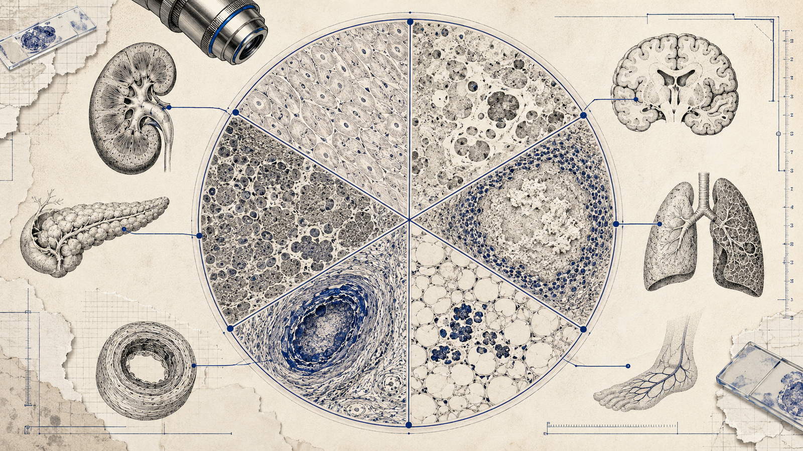
| Coagulative | Ischemic injury in most solid organs (e.g., myocardial infarction, renal infarction) — architecture is preserved for a time |
| Liquefactive | Brain infarction and bacterial abscesses — enzymatic digestion dissolves tissue into liquid |
| Caseous | Tuberculosis — a distinctive combination of coagulative and liquefactive features |
| Fat | Acute pancreatitis, and traumatic fat necrosis in the breast |
| Fibrinoid | Immune-mediated vascular damage — malignant hypertension, vasculitis, and the Aschoff bodies of rheumatic fever |
| Gangrenous | Ischemic limb or bowel — essentially coagulative necrosis, with a "wet" form when infection is superimposed |
This table is the single highest-yield asset in the whole topic — a described finding maps to a type, and a type maps to a diagnosis, in that order.
Coagulative Necrosis: Why Architecture Survives, For a While
Coagulative necrosis is the default pattern of necrosis in most solid organs following ischemia — the heart, kidney, and spleen are the classic examples. The defining feature is that the tissue's basic architecture is preserved for several days after cell death, even though the cells themselves are dead. This happens because the ischemic insult denatures both structural proteins and the enzymes that would otherwise digest the tissue — without functional proteolytic enzymes, the dead cells retain their outline even as they lose function entirely. A myocardial infarction is the textbook example: on histology, you can still make out the ghost outlines of cardiac myocytes days after the infarct, before the tissue is eventually cleared by macrophages and replaced with scar.
Liquefactive Necrosis: When Enzymes Win
Liquefactive necrosis is what happens when proteolytic enzymes are NOT denatured alongside the structural proteins — the enzymes remain active and digest the dead tissue into a liquid, viscous mass. The two classic settings are brain infarction and bacterial infection (abscess formation). The brain is a special case worth understanding, not just memorizing: brain tissue is lipid-rich and enzyme-rich relative to other organs, which makes it prone to enzymatic autolysis even after ischemic injury that would produce coagulative necrosis anywhere else in the body. In bacterial infections, the liquefaction is driven by enzymes released from the massive numbers of neutrophils recruited to fight the infection — which is exactly why an abscess is, physically, a liquefied collection of dead tissue and inflammatory cells.
Caseous Necrosis: A Named Hybrid, Not a Third Mechanism
Caseous necrosis is worth understanding as a distinctive combination of the two mechanisms above, rather than a wholly separate process — it has the appearance of coagulative necrosis (structureless, without liquefaction) but with a granular, "cheese-like" quality that doesn't fit cleanly into either coagulative or liquefactive on its own. It's classically associated with tuberculosis, and on histology it's typically surrounded by a granulomatous inflammatory response — a ring of activated macrophages (epithelioid cells), giant cells, and lymphocytes. Recognizing caseous necrosis on a description is often the fastest route to a tuberculosis diagnosis in a vignette, since few other conditions produce this specific combination of findings.
Fat Necrosis: Two Different Mechanisms, Same Name
Fat necrosis is worth separating into two genuinely distinct clinical scenarios that happen to share a name. Enzymatic fat necrosis occurs in acute pancreatitis, where released pancreatic lipases digest peripancreatic and intra-abdominal fat — the released fatty acids then combine with calcium to form visible, chalky-white calcium soaps, which is why hypocalcemia can be a real clinical complication of severe pancreatitis (calcium gets consumed in this reaction). Traumatic fat necrosis occurs in fat-rich tissue, most classically the breast, following blunt trauma — it can form a firm, sometimes painless mass that is a genuine diagnostic mimic of breast cancer on examination and imaging, which is exactly why it matters clinically as well as on the exam.
Fibrinoid Necrosis: A Vascular, Immune-Mediated Pattern
Fibrinoid necrosis is distinct from the others in both mechanism and location — it occurs specifically within blood vessel walls, and it's immune-mediated rather than purely ischemic. Immune complexes deposit in the vessel wall alongside fibrin, producing a bright pink, amorphous appearance on histology that resembles fibrin under the microscope (hence the name). The classic contexts are malignant hypertension (where the acutely elevated pressure itself damages small vessel walls), vasculitis (where immune-mediated vessel wall inflammation is the primary process), and the Aschoff bodies of rheumatic fever, which contain a core of fibrinoid necrosis surrounded by a specific inflammatory cell population. Recognizing fibrinoid necrosis as vessel-specific and immune-mediated — rather than a generic ischemic pattern — is what separates it from the other five types.
Gangrenous Necrosis: Not a Separate Mechanism, But a Useful Clinical Term
Gangrenous necrosis is, mechanistically, coagulative necrosis affecting an extremity or the bowel following ischemia — it's a clinical/anatomical term more than a distinct histological pattern. The dry-versus-wet distinction is the exam-relevant detail: dry gangrene is the coagulative pattern alone, with the tissue becoming dry, shrunken, and darkened as blood supply is lost. Wet gangrene occurs when bacterial infection is superimposed on the ischemic, coagulative tissue, adding a liquefactive component on top — the tissue becomes swollen, foul-smelling, and the infection can spread rapidly, making wet gangrene a genuine surgical emergency in a way dry gangrene, while serious, typically isn't to the same degree.
Necrosis vs Apoptosis: The Distinction Worth Keeping Sharp
Necrosis and apoptosis are both forms of cell death, but they diverge in trigger, mechanism, and downstream consequence. Necrosis is typically pathological — triggered by an external insult like ischemia, toxins, or trauma — and provokes inflammation, since cell membrane breakdown spills contents into surrounding tissue. Apoptosis is a programmed, energy-dependent process, often physiological (normal tissue turnover, embryological development) but sometimes pathological too (elimination of a virally infected cell), and it characteristically does not provoke inflammation, since the cell fragments into membrane-bound apoptotic bodies that are cleared without spilling contents. A question describing inflammation accompanying cell death is pointing toward necrosis; one describing an isolated cell dying without a surrounding tissue reaction is pointing toward apoptosis.
A Few Pairings Worth Locking In
- Caseous necrosis + granulomatous inflammation almost always means tuberculosis (or occasionally other granulomatous infections) — the pairing is more diagnostic than either finding alone.
- Fat necrosis + hypocalcemia in a patient with acute abdominal pain points toward severe pancreatitis, via the calcium-soap mechanism.
- Fibrinoid necrosis should immediately narrow your differential to vessel-wall pathology — malignant hypertension, vasculitis, or rheumatic fever — not a generic ischemic process.
- Liquefactive necrosis in the brain specifically doesn't require an infection to explain it — ischemic stroke alone produces this pattern in neural tissue, unlike almost anywhere else in the body.
For the broader pattern this kind of question rewards, our guide to anemia classification covers the same lab-and-morphology recognition skill applied to a different system, and our subject-wise weightage breakdown covers how much of the paper Pathology carries overall.
Practise this on Reflex
Turn what you just read into recall with 14 years of tagged PYQs.
FAQ
Frequently asked questions
The questions aspirants ask most about this topic.
Coagulative necrosis — the classic pattern in most solid organs following ischemia, with cellular architecture preserved for several days after cell death.
Brain tissue is lipid-rich and enzyme-rich relative to other organs, making it prone to enzymatic autolysis after ischemic injury — a pattern that would produce coagulative necrosis anywhere else in the body.
In acute pancreatitis, released pancreatic lipases digest peripancreatic fat, and the resulting fatty acids combine with calcium to form visible, chalky-white deposits — a reaction that can also contribute to clinically significant hypocalcemia.
Dry gangrene is coagulative necrosis alone, from ischemia. Wet gangrene adds a liquefactive component from superimposed bacterial infection, making it a more urgent surgical emergency.
Caseous necrosis has a granular, "cheese-like" quality that doesn't fully liquefy the way liquefactive necrosis does, but also doesn't preserve architecture as cleanly as pure coagulative necrosis — it's classically associated with tuberculosis and typically surrounded by granulomatous inflammation.
About the author
Stay ahead in your preparation
Get expert tips, exam updates, and high-yield insights delivered straight to your inbox.
No spam. Unsubscribe anytime.
More in Pathology
View all → Pathology
Types of Hypersensitivity Reactions: The Gell and Coombs Classification NEET PG Tests Every Cycle
 Pathology
Reed-Sternberg Cells and Hodgkin vs Non-Hodgkin Lymphoma: The Full NEET PG Comparison
 Pathology
Tumor Markers for NEET PG: The Complete List and What They Actually Mean
Every high-yield marker matched to its cancer, plus the distinction examiners love: monitoring vs screening.
 Pathology
How to Prepare for NEET PG/INICET Pathology: A Complete Guide
Pathology is not just another subject. It forms a core scoring pillar in NEET PG/INICET.