Types of Hypersensitivity Reactions: The Gell and Coombs Classification NEET PG Tests Every Cycle
By Dr. Utsav Bhattacherjee, MBBS, MBA · 4 Sept 2026 · 11 min read
Last updated: 23 Sept 2026
On this page
Hypersensitivity reaction questions are built entirely around the Gell and Coombs classification — four types, each defined by a different immune mechanism, not just a different clinical picture. Learn the mechanism behind each type, and the examples stop being a memorized list and start being predictable consequences of that mechanism.
Type I: Immediate (Anaphylactic) Hypersensitivity
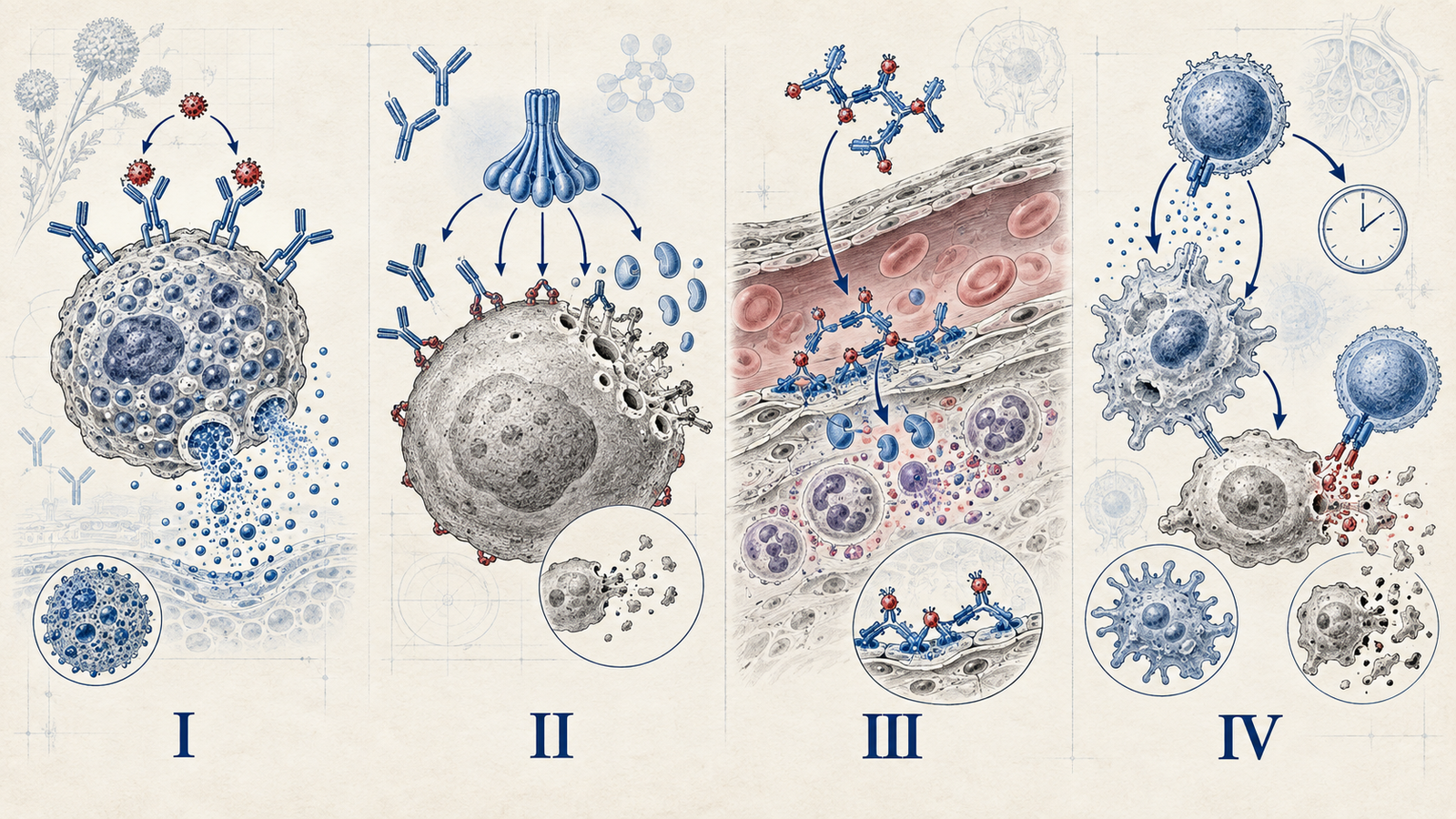
Type I is IgE-mediated. On first exposure to an allergen, the immune system produces IgE antibodies specific to it, which then bind to receptors on mast cells and basophils — this is sensitization, and it produces no symptoms on its own. On re-exposure, the allergen cross-links the IgE already bound to mast cells, triggering immediate degranulation — release of histamine and other mediators — which is why Type I reactions happen within minutes of re-exposure, not on first contact with the allergen.
Classic examples: anaphylaxis, allergic asthma, allergic rhinitis (hay fever), urticaria, and food allergies. The speed of onset — minutes, not hours or days — is itself a diagnostic clue distinguishing Type I from the other three types.
Type II: Cytotoxic Hypersensitivity
Type II involves IgG or IgM antibodies directed against antigens on the surface of the body's own cells. Once bound, these antibodies can destroy the target cell through several mechanisms: activating the complement cascade directly, or flagging the cell for destruction by phagocytes or NK cells (antibody-dependent cellular cytotoxicity).
Classic examples: hemolytic disease of the newborn (maternal antibodies against fetal Rh antigen), Goodpasture syndrome (antibodies against glomerular and alveolar basement membrane), autoimmune hemolytic anemia, and Rheumatic fever's molecular mimicry against cardiac tissue.
The exam-favorite exception worth knowing separately: some conditions classified under Type II don't destroy cells at all — they work through antibodies binding receptors and either stimulating or blocking them, without cytotoxicity. Graves' disease (antibodies stimulating the TSH receptor) and myasthenia gravis (antibodies blocking the acetylcholine receptor) are both classified as Type II despite involving no cell destruction whatsoever — a detail that catches students who assume Type II always means cytotoxicity.
Type III: Immune Complex Hypersensitivity
Type III involves antigen-antibody complexes that form in circulation and then deposit in tissues — blood vessel walls, joints, kidneys, and skin are classic deposition sites. Once deposited, these complexes activate complement, and the resulting inflammatory response is what actually causes tissue damage — the damage is a bystander effect of complexes lodging somewhere, not a direct, targeted attack on that specific tissue the way Type II is.
Classic examples: systemic lupus erythematosus (complexes depositing in kidneys, joints, and skin), serum sickness (a classic reaction to foreign protein, such as antitoxin), post-streptococcal glomerulonephritis, and the Arthus reaction (a localized version, classically demonstrated at an injection site).
Type IV: Delayed-Type (Cell-Mediated) Hypersensitivity
Type IV is the outlier of the four in one crucial respect: it involves no antibodies at all. It's mediated entirely by T cells — sensitized T cells recognizing an antigen and recruiting macrophages and other inflammatory cells to the site, a process that takes real time to unfold. This is exactly why Type IV reactions are delayed, classically appearing 24–72 hours after exposure, in sharp contrast to Type I's minutes-scale onset.
Classic examples: the tuberculin (PPD/Mantoux) skin test reaction, contact dermatitis (poison ivy being the classic trigger), graft rejection, and the autoimmune destruction of pancreatic beta cells in Type 1 diabetes.
The Four Types, Side by Side
| Type | Mechanism | Onset | Classic Example |
|---|---|---|---|
| I | IgE, mast cell degranulation | Minutes | Anaphylaxis |
| II | IgG/IgM against cell-surface antigens | Hours | Hemolytic disease of newborn |
| III | Immune complex deposition | Hours to days | SLE, serum sickness |
| IV | T-cell mediated, no antibody | 24–72 hours | TB skin test, contact dermatitis |
Why the Type II Receptor Exception Matters So Much on Exams
Graves' disease and myasthenia gravis are worth dwelling on specifically because they're the detail most likely to trip up an otherwise solid understanding of this topic. If your working definition of Type II is "antibody destroys a cell," both of these conditions will seem to not fit — no cell is being destroyed in either case. The broader, correct definition is "antibody directed against a cell-surface antigen," which covers destruction and receptor stimulation or blockade as two different downstream consequences of the same underlying mechanism. Holding onto the broader definition rather than the narrower "cytotoxic" shorthand is what actually prevents this specific exam trap.
FAQs
What is the main difference between Type II and Type III hypersensitivity?
Type II antibodies target antigens directly on the surface of a specific cell, causing localized damage to that cell type. Type III involves antigen-antibody complexes that form in circulation and then deposit wherever they happen to lodge, causing damage as a bystander effect rather than a targeted attack on one cell type.
Why are Graves' disease and myasthenia gravis classified as Type II if no cells are destroyed?
Type II is defined by antibodies targeting cell-surface antigens, not exclusively by cell destruction. In these two conditions, antibodies bind and either stimulate (Graves') or block (myasthenia gravis) a receptor without destroying the cell itself, which still fits the broader Type II mechanism.
Why is Type IV hypersensitivity called "delayed"?
Because it's T-cell mediated rather than antibody-mediated, and recruiting and activating T cells and macrophages at the exposure site takes real time — classically 24 to 72 hours — unlike the minutes-scale onset of antibody-driven Type I reactions.
Which hypersensitivity type is responsible for the tuberculin skin test reaction?
Type IV. The reaction to PPD is a classic example of delayed-type, T-cell mediated hypersensitivity, which is exactly why the test is read 48–72 hours after placement rather than immediately.
What role does complement play in Type III hypersensitivity?
Once immune complexes deposit in tissue, they activate the complement cascade, and the resulting inflammatory response is what actually causes the tissue damage seen in conditions like lupus or serum sickness.
For the same mechanism-first approach applied to a different immune-mediated topic, see our guide to nephrotic vs nephritic syndrome, and for how much of the paper Pathology carries overall, our subject-wise weightage breakdown.
Practise this on Reflex
Turn what you just read into recall with 14 years of tagged PYQs.
FAQ
Frequently asked questions
The questions aspirants ask most about this topic.
Type II antibodies target antigens directly on the surface of a specific cell, causing localized damage to that cell type. Type III involves antigen-antibody complexes that form in circulation and deposit wherever they happen to lodge, causing damage as a bystander effect.
Type II is defined by antibodies targeting cell-surface antigens, not exclusively by cell destruction. In these two conditions, antibodies bind and either stimulate or block a receptor without destroying the cell itself.
Because it's T-cell mediated rather than antibody-mediated, and recruiting and activating T cells and macrophages at the exposure site takes real time — classically 24 to 72 hours.
Type IV. The reaction to PPD is a classic example of delayed-type, T-cell mediated hypersensitivity, which is why the test is read 48–72 hours after placement.
Once immune complexes deposit in tissue, they activate the complement cascade, and the resulting inflammatory response is what causes the tissue damage seen in conditions like lupus or serum sickness.
About the author
Stay ahead in your preparation
Get expert tips, exam updates, and high-yield insights delivered straight to your inbox.
No spam. Unsubscribe anytime.
More in Pathology
View all → Pathology
Reed-Sternberg Cells and Hodgkin vs Non-Hodgkin Lymphoma: The Full NEET PG Comparison
 Pathology
Types of Necrosis for NEET PG: The Pattern-to-Diagnosis Table Examiners Actually Test
 Pathology
Tumor Markers for NEET PG: The Complete List and What They Actually Mean
Every high-yield marker matched to its cancer, plus the distinction examiners love: monitoring vs screening.
 Pathology
How to Prepare for NEET PG/INICET Pathology: A Complete Guide
Pathology is not just another subject. It forms a core scoring pillar in NEET PG/INICET.