Amyloidosis for NEET PG: Congo Red, Apple-Green Birefringence and the Types of Amyloid Protein
Reflex · 18 Nov 2026 · 11 min read
Last updated: 18 Nov 2026
On this page
Amyloidosis is a pathology topic where one test finds the disease and another names it. Congo red staining with apple-green birefringence under polarised light shows that amyloid is present. It cannot say whether the deposit is AL, AA, ATTR or another type, and that distinction decides treatment.
Key Takeaways
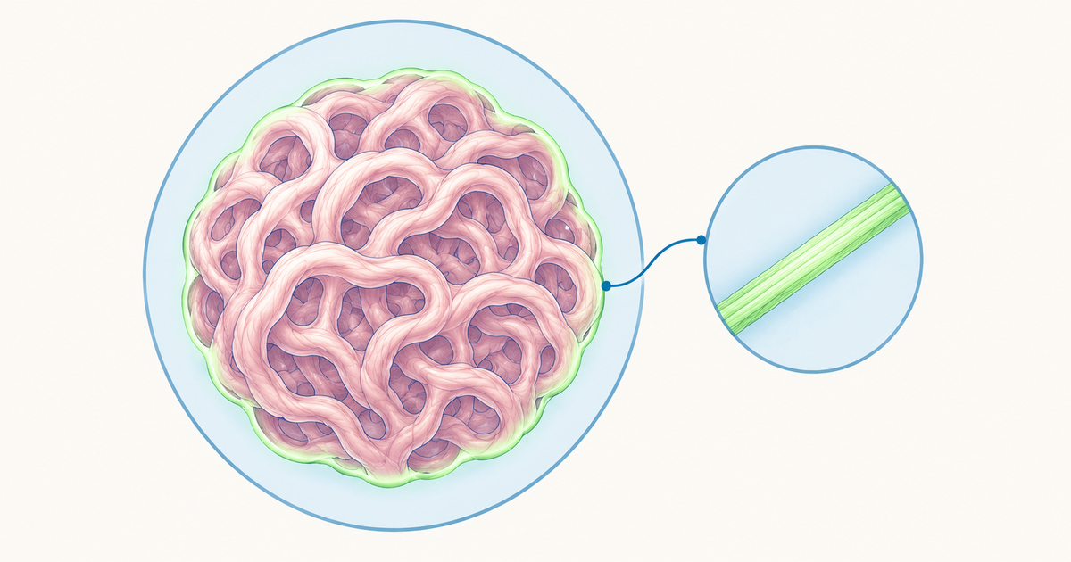
Amyloid is extracellular protein misfolded into insoluble cross-beta sheet fibrils.
Congo red looks orange-red or salmon-pink in ordinary light and apple-green under polarised light. Real slides can look yellow-green or blue-green, and a negative stain does not exclude amyloid.
Congo red confirms amyloid but never names the type. Typing needs immunohistochemistry or laser microdissection with mass spectrometry.
Amyloid is named "A" plus its precursor: AL (light chain), AA (serum amyloid A), ATTR (transthyretin), Abeta2M (beta-2 microglobulin, long-term dialysis).
AL arises from a clonal plasma cell or B-cell disorder and AA from chronic inflammation, so treatment targets the clone or the inflammation.
Screen for AL with serum and urine immunofixation plus serum free light chains, not electrophoresis alone.
What Is Amyloidosis?
Amyloidosis is a group of diseases in which misfolded protein deposits as insoluble fibrils in tissues and alters their function (American Society of Hematology (ASH) 2026 guideline). The deposits are extracellular, and the protein's native structure changes to a predominantly cross-beta sheet form (DES-Amyloidoses knowledgebase). Within the cross-beta structure, the beta-strands stack perpendicular to the fibril axis, and the fibrils resist proteases and detergents and usually bind Congo red and thioflavin T (renal diagnostics review).
Deposits also contain proteoglycans, glycosaminoglycans, serum amyloid P component (SAP) and apolipoproteins. SAP binds the fibrils and helps them resist breakdown (proteomics review).
How Amyloid Is Named
The name is "A" for amyloid followed by an abbreviation of the precursor protein, so immunoglobulin light chain deposits are AL. Hereditary forms add the variant, as in ATTR V30M (proteomics review). The older labels "primary", "secondary" and "hereditary" date from 1970s classification systems and have been replaced by protein-based naming (ASH guideline).
The ASH guideline counts 42 human amyloid fibril proteins, as does the International Society of Amyloidosis (ISA) 2024 nomenclature report, and the number has risen over time (DES-Amyloidoses knowledgebase). The cell injury notes cover the wider picture of tissue injury.
Memory aid: read every name as "A + the precursor". AL is Light chain, AA is Acute-phase serum amyloid A, ATTR is TransThyRetin, and Abeta2M is Beta-2 Microglobulin.
Congo Red and Apple-Green Birefringence
What the Stain Shows
Positive Congo red staining with apple-green birefringence under polarised light is the gold standard for diagnosing amyloid in tissue (renal diagnostics review). In ordinary bright-field light, stained amyloid appears orange-red (proteomics review) or salmon-pink (Kidney360 review). Under crossed polarisers it shows apple-green birefringence (Congo red review).
Memory aid: red on the slide, green under the polarisers.
Why Green Is the Textbook Colour but Not the Whole Story
Across 160 papers on Congo red staining in tissue sections, only 31% of the published images showed a pure green colour. Yellow-green and blue-green are common in practice, and the same area of a section can look green or yellow depending on microscope setup (Congo red review). Learn "apple-green" as the textbook description, not a pure-green rule.
Why Congo Red Alone Cannot Name the Type
Fibrils of every amyloid type have the cross-beta structure that usually binds Congo red. Apple-green birefringence is the textbook description of the stained deposit, so a positive stain confirms amyloid without naming the protein (renal diagnostics review). After amyloid is established, the precursor protein must be identified in a separate step (Kidney360 review).
Overstaining can give false positives (proteomics review), and the dye also stains collagen, elastic fibres and hyaline deposits (Congo red review). That review concluded that Congo red alone is insufficient for diagnosing amyloidosis, and a negative result should prompt immunohistochemistry (IHC).
Sensitivity depends on the tissue sampled (Congo red review), and fat-pad aspirates range from 13% to 73% across studies (gastrointestinal review). A negative stain does not exclude amyloid (Kidney360 review).
Other Ways to Detect and Identify Amyloid
| Method | What it shows | Role |
|---|---|---|
| Thioflavin T | Strong fluorescence on binding; also stains cartilage and elastic fibres | More sensitive than Congo red, but used mainly in research in renal pathology |
| Electron microscopy | Unbranched, randomly arranged fibrils 7-12 nm in diameter | Helps when Congo red is equivocal |
| X-ray diffraction | Cross-beta pattern: strands perpendicular to the fibril axis (4.7 Å spacing), sheets about 10 Å apart | A research technique, not a routine clinical test |
| IHC or immunofluorescence | Antibodies against kappa and lambda light chains, serum amyloid A (SAA), transthyretin (TTR) and others | Common typing method |
| Laser microdissection with mass spectrometry (LMD-MS) | Sequences the proteins in the Congo red-positive area | Typing method regarded as the gold standard |
Sources: the Congo red review, the Kidney360 review and the renal diagnostics review.
Types of Amyloid Protein: Classification of Amyloidosis
Amyloidosis is classified by the fibril protein, then split into systemic types (deposits from circulating precursors) and localised types (deposits from local production). Systemic amyloidoses carry a worse prognosis than localised forms (renal diagnostics review).
| Type | Precursor protein | Pattern | Main sites or setting |
|---|---|---|---|
| AL | Immunoglobulin light chain (kappa or lambda) | Systemic and localised | Clonal plasma cell or B-cell disorder; heart, kidneys, gastrointestinal (GI) tract, peripheral nerves |
| AA | Serum amyloid A (acute-phase protein) | Systemic | Chronic inflammation or infection; kidney and GI tract |
| ATTR wild-type ("senile") | Normal transthyretin | Systemic | Heart mainly; also lung, ligaments and tenosynovium |
| ATTR hereditary (ATTRv) | Variant transthyretin | Systemic | Heart, peripheral and autonomic nerves, eye; mutations include Val30Met, Val122Ile and Thr60Ala |
| Abeta2M | Beta-2 microglobulin | Systemic | Long-term haemodialysis; bones and ligaments |
| ALECT2 | Leukocyte chemotactic factor 2 | Systemic | Kidney, mainly in older people; more frequent in Hispanic populations |
| AFib | Fibrinogen A-alpha variants | Systemic, hereditary | Kidney, primarily |
| AApoAI | Apolipoprotein A-I variants | Systemic, hereditary | Heart, liver, kidney, peripheral nerves, testis; larynx and skin for C-terminal variants |
| AApoAII, ALys | Apolipoprotein A-II and lysozyme variants | Systemic, hereditary | Kidney |
| AGel | Gelsolin variants | Systemic, hereditary | Kidney, peripheral nerves, cornea |
| ACys | Cystatin C variants | Systemic, hereditary | Peripheral nerves, skin; central nervous system (CNS) in the open-access table |
| Abeta | Abeta protein precursor | Localised | Brain (CNS); Alzheimer's disease plaques |
| ACal | (Pro)calcitonin | Localised; a systemic form is also listed | C-cell thyroid tumours (medullary thyroid carcinoma); kidney in the systemic form |
| AIAPP | Islet amyloid polypeptide (amylin) | Localised | Islets of Langerhans; main component of islet amyloid in type 2 diabetes |
| AANP (older name AANF) | Atrial natriuretic peptide | Localised | Cardiac atria |
| AIns | Insulin | Localised | Injection sites |
| APro | Prolactin | Localised | Pituitary prolactinomas and the ageing pituitary |
Primary source: the ISA 2024 report. Open-access check: a 2022 review table adapted from an earlier ISA nomenclature update.
The ISA report classes ATTR as systemic but says it is not fully answered whether ATTR can appear as a strictly localised form.
Cited for the table: AL organs (ASH guideline); AA, ATTR and ALECT2 organs, ATTR mutations, Hispanic predominance (renal diagnostics review); AA GI involvement (gastrointestinal review); haemodialysis (beta2-microglobulin review); type 2 diabetes (islet amyloid polypeptide review).
AL vs AA Amyloid: The Contrast That Matters
A positive Congo red slide does not separate AL from AA. AL fibrils come from immunoglobulin light chains made by a clonal plasma cell or B-cell population (renal diagnostics review), although only a minority of patients have a true neoplasm (proteomics review). AA fibrils come from serum amyloid A, an acute-phase protein made in response to chronic inflammation or infection (gastrointestinal review, proteomics review).
| Feature | AL | AA | ATTR |
|---|---|---|---|
| Fibril protein | Immunoglobulin light chain | Serum amyloid A | Transthyretin |
| Setting | Clonal plasma cell or B-cell disorder (a true neoplasm in a minority) | Chronic inflammation or infection | Hereditary variant or age-related wild type |
| Main organs | Heart, kidney, GI tract, peripheral nerves | Kidney; highest rate of GI involvement | Heart mainly; nerves in the hereditary form |
| Typing test | IHC for kappa and lambda, or LMD-MS | Anti-SAA IHC or LMD-MS | Anti-TTR IHC; genetics for hereditary disease |
Organs row: AL (ASH guideline); AA (renal diagnostics review, gastrointestinal review); ATTR (Kidney360 review).
AL is the most common systemic type in developed countries, while triggers for AA include rheumatoid arthritis, Crohn's disease, chronic osteomyelitis and bronchiectasis (renal diagnostics review).
How Amyloidosis Is Diagnosed
The diagnostic path has three steps:
- Suspect it: clues and monoclonal protein screening.
- Prove it in tissue: Congo red-stained biopsy.
- Type it: IHC or LMD-MS.
The screening and biopsy advice below is for suspected AL amyloidosis, the most common systemic type in developed countries (renal diagnostics review) and the subject of the 2026 ASH guideline.
Step 1: Suspect It
Suspicion rises with nephrotic syndrome, unexplained cardiac dysfunction, autonomic or sensory neuropathy, periorbital purpura and weight loss; renal amyloid usually presents with nephrotic syndrome and chronic kidney disease (CKD) (renal diagnostics review). The nephrotic vs nephritic syndrome notes add background.
For suspected cardiac amyloidosis (and, conditionally, unexplained proteinuria), ASH recommends serum and urine immunofixation (SIFE and UIFE) plus serum free light chains (sFLC) with their ratio. Suspicion is enhanced if any one test is positive. Serum and urine protein electrophoresis (SPEP and UPEP) should not be the screening tests or the only tests (ASH guideline).
In a study of 115 patients with monoclonal gammopathy, the sFLC ratio alone had 76% sensitivity, while immunofixation identified the amyloidogenic light chain in all cases. These tests are not specific, because monoclonal gammopathy of undetermined significance (MGUS) is far more common than AL (renal diagnostics review).
Step 2: Prove It With Tissue
AL is confirmed by tissue analysis. A surrogate biopsy is a less invasive substitute for a target-organ biopsy, most often from abdominal fat pad or bone marrow. The target organs are the heart, kidneys, liver and peripheral nerves, and enough tissue must be collected to allow subtyping (ASH guideline).
ASH makes conditional recommendations by suspected organ:
- Suspected renal AL with positive paraprotein tests: fat-pad plus bone marrow biopsy before renal biopsy, which a positive subtyped result makes unnecessary.
- Suspected cardiac AL: fat-pad plus bone marrow biopsy, or endomyocardial biopsy, which may be favoured when hereditary ATTR risk factors exist.
Liver biopsy is discouraged when tissue can be obtained elsewhere, because of haemorrhage risk (gastrointestinal review).
Step 3: Type It
ASH states as good practice that once amyloid is proven on histology, subtyping with a validated method must follow promptly (ASH guideline).
- IHC or immunofluorescence: the most common typing method in clinical laboratories, because it is cheaper and more reproducible. Results depend on reagent quality and pathologist experience, with a false-negative risk in AL and a false-positive risk in AA (renal diagnostics review). Commercial antibodies miss the mutated light chain in 10-15% of AL patients (Kidney360 review).
- LMD-MS: laser microdissection of the Congo red-positive area, then tandem mass spectrometry. Studies report typing in about 92-98% of cases for LMD-MS against about 42-45% for IHC (renal diagnostics review, proteomics review).
Imaging Tests
Iodine-123-labelled SAP scintigraphy can show that amyloid is present but cannot identify the type (Kidney360 review). Bone scintigraphy with technetium-99m-labelled pyrophosphate or diphosphonate tracers is different. ASH strongly recommends against it for diagnosing AL cardiac amyloidosis. It strongly recommends it, with single-photon emission computed tomography (SPECT), for cardiac ATTR when there is no plasma cell disorder (ASH guideline).
The reason is overlap: plasma cell dyscrasia was found in 39% of wild-type and 49% of hereditary ATTR cases, so paraprotein testing comes first (renal diagnostics review).
Treatment Follows the Amyloid Type
Typing matters because treatments differ (renal diagnostics review). This outline is for orientation, and ASH recommendations are written for high-income settings (ASH guideline).
- AL: treat the plasma cell clone. First-line treatment includes D-VCD (daratumumab, bortezomib, cyclophosphamide, dexamethasone) and, in some patients, high-dose chemotherapy with autologous haematopoietic cell transplantation (ASH guideline).
- AA: suppress the underlying inflammation. Lowering serum amyloid A below 10 mg/L was associated with amyloid regression and better survival. In familial Mediterranean fever, colchicine reduced the prevalence of nephropathy (Kidney360 review).
- Hereditary ATTR: liver transplantation (the liver is the main source of transthyretin), TTR tetramer stabilisers, and antisense or TTR-mRNA-silencing agents, which were effective in phase 3 trials (renal diagnostics review).
Revising Amyloidosis: Study Tips and Common Mistakes
Study Tips
- Draw the AL vs AA vs ATTR table from memory, then practise with the pathology practice questions and use the pathology preparation guide for the wider syllabus.
- If a stem gives rheumatoid arthritis, chronic osteomyelitis or bronchiectasis with proteinuria, think AA. If it gives myeloma or a monoclonal protein, think AL. If it gives long-term haemodialysis, think Abeta2M.
- Test yourself: two biopsies are Congo red positive, one from a patient with long-standing rheumatoid arthritis and one with a monoclonal protein. What is the next step? Type both, because the stain confirms amyloid but cannot separate AA from AL.
Common Mistakes to Avoid
- Insisting on pure green birefringence, or treating a negative Congo red as proof that amyloid is absent.
- Using SPEP or UPEP alone to screen for AL instead of immunofixation plus sFLC.
- Choosing bone scintigraphy to diagnose AL cardiac amyloid. It is for cardiac ATTR once a plasma cell disorder is excluded.
- Using "primary" and "secondary" as if they were current names rather than the fibril protein.
Amyloidosis Revision Summary
Congo red with apple-green birefringence shows that amyloid is present, and typing shows which protein it is. Because the types differ in cause, organs and treatment, keep the AL vs AA contrast, the classification table and the three diagnostic steps.
Practise this on Reflex
Turn what you just read into recall with 14 years of tagged PYQs.
FAQ
Frequently asked questions
The questions aspirants ask most about this topic.
Congo red shows that amyloid is present but not which protein forms it. Fibrils of every amyloid type have the cross-beta structure that usually binds the dye, and apple-green birefringence is the textbook description of the stained deposit, so the precursor protein is identified in a separate typing step. A 2019 review concluded that Congo red alone is insufficient for diagnosing amyloidosis, and the type is assigned by immunohistochemistry or by laser microdissection with mass spectrometry.
No. Apple-green is the textbook description of Congo red-stained amyloid under polarised light, but in a review of 160 papers only 31% of the published images showed a pure green colour. Yellow-green and blue-green appear in practice, and the same area of a section can look green or yellow depending on the microscope setup.
AL amyloid is made of immunoglobulin light chains from a clonal plasma cell or B-cell disorder and mainly affects the heart, kidneys, gastrointestinal (GI) tract and peripheral nerves. AA amyloid is made of serum amyloid A, an acute-phase protein raised in chronic inflammation or infection, and mainly affects the kidney and GI tract. A positive Congo red result does not separate them, so typing does, and treatment follows the type: the plasma cell clone in AL and the underlying inflammation in AA.
The 2026 American Society of Hematology (ASH) guideline on AL amyloidosis strongly recommends serum and urine immunofixation plus serum free light chains with their ratio to raise suspicion when cardiac amyloidosis is suspected, conditionally suggests the same panel for unexplained proteinuria, and states as good practice that serum and urine protein electrophoresis should not be the screening tests or the only tests. In one study of 115 patients with monoclonal gammopathy, immunofixation identified the amyloidogenic light chain in all of them, while the free light chain ratio alone had 76% sensitivity.
Laser microdissection of the Congo red-positive area followed by tandem mass spectrometry (LMD-MS) is described as the gold standard for typing. Studies report typing in about 92-98% of cases with LMD-MS against about 42-45% with immunohistochemistry. Immunoassays remain more widely used in clinical laboratories because they are cheaper and more reproducible.
Stay ahead in your preparation
Get expert tips, exam updates, and high-yield insights delivered straight to your inbox.
No spam. Unsubscribe anytime.
More in Pathology
View all → Pathology
Cell Injury for NEET PG: Reversible vs Irreversible Changes and the Point of No Return
A structured walk through cell injury: what causes it, how ATP loss and calcium shifts drive it, how to tell reversible from irreversible change under the microscope, and why the point of no return is not one single event.
 Pathology
Virchow's Triad for NEET PG: Three Components, Each With Clinical Examples
Virchow's triad lists the three factors that promote thrombosis: endothelial injury, abnormal blood flow and hypercoagulability. Learn each component with clinical examples and a worked renal cell carcinoma case.
 Pathology
Types of Hypersensitivity Reactions: The Gell and Coombs Classification NEET PG Tests Every Cycle
 Pathology