Virchow's Triad for NEET PG: Three Components, Each With Clinical Examples
Reflex · 29 Oct 2026 · 10 min read
Last updated: 29 Oct 2026
On this page
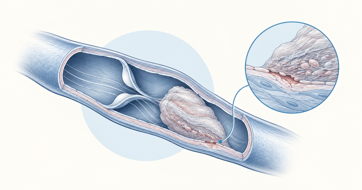
Virchow's triad is the standard framework for explaining why a thrombus forms inside an intact circulation. It names three factors: injury to the vessel wall, abnormal blood flow, and a blood that clots too readily. Every thrombosis question in pathology, from deep vein thrombosis to a clot on a ruptured plaque, can be reduced to which of these three is doing the work.
This post takes each component in turn, gives the clinical examples worth remembering, separates inherited from acquired hypercoagulability, and then applies all three to one patient. It also clears up two points that confuse students: who actually proposed the triad, and how it differs from the Virchow node.
Key Takeaways
Virchow's triad has three components: endothelial injury, abnormal blood flow (stasis or turbulence), and hypercoagulability.
Thrombosis is believed to need at least two of the three factors acting together, and the usual example lists are not exhaustive.
Endothelial injury covers plaque rupture, vasculitis, valve disease, endotoxins and catheters. Stasis covers immobility, varicose veins, heart failure and aneurysm.
Hypercoagulability splits into inherited causes (factor V Leiden, protein C, protein S and antithrombin deficiency) and acquired causes (malignancy, pregnancy, oral contraceptives, antiphospholipid syndrome, nephrotic syndrome).
Renal cell carcinoma shows all three components in one patient.
The triad is named after Rudolf Virchow, but the triad as a formal list was not his. The Virchow node is a separate eponym.
What Is Virchow's Triad?
Virchow's triad is a list of three factors that promote thrombosis: endothelial injury, disturbance of blood flow (slow or turbulent), and hypercoagulability. Radiopaedia describes it as the three broad categories of factors believed to contribute to thrombosis, and notes that it is thought to take at least two of them together to cause a clot. The examples under each heading are illustrative, not complete.
That "at least two" idea is useful in practice. A single risk factor, such as a long flight, rarely tells the whole story. When a patient develops a clot, ask which other component was also present.
A simple memory aid is Wall, Flow, Blood:
- Wall: the endothelium is damaged.
- Flow: blood moves too slowly or too turbulently.
- Blood: the blood itself is prone to clot.
Virchow Triad at a Glance
The table below is the one to revise the night before a test. Each row gives the component, what it does to coagulation, and the examples that appear most reliably across references.
| Component | What goes wrong | Clinical examples |
|---|---|---|
| Endothelial injury | The vessel lining is damaged; in plaque rupture, exposed tissue factor activates coagulation and platelets adhere | Ulcerated atheromatous plaque, myocardial infarction, valvular heart disease, vasculitis, endotoxins and sepsis, trauma or surgery, venous catheters |
| Abnormal blood flow | Flow is slow (stasis) or disorganised (turbulence), which promotes clotting | Immobility and bed rest, varicose veins, heart failure, arterial aneurysm, pregnancy, obesity, long travel |
| Hypercoagulability | The blood has an increased tendency to clot, from a gene change or an acquired condition | Factor V Leiden, protein C, protein S or antithrombin deficiency, malignancy, oral contraceptives, antiphospholipid syndrome, nephrotic syndrome |
Sources list these examples as non-exhaustive, so treat any single option in a question stem as a clue to the dominant component rather than a complete explanation.
Endothelial Injury in Virchow's Triad
Endothelial injury is the first component: damage to the vessel lining exposes the tissue beneath it. In arteries, the classic event is atherosclerotic plaque rupture. The exposed tissue factor activates coagulation and triggers platelet adhesion and aggregation, as described in the MSD Manual.
Clinical examples of endothelial injury
Students tend to remember plaque rupture and forget the rest. These are the examples worth holding together:
- Ulcerated atheromatous plaques, which explain arterial thrombosis.
- Myocardial infarction, listed by Radiopaedia among the causes of endothelial injury.
- Cardiac valve disease, also listed by Radiopaedia under endothelial injury.
- Vasculitis, where inflamed vessel walls lose their normal surface.
- Endotoxins and severe infection: in sepsis, monocytes and macrophages express more tissue factor.
- Tissue injury from trauma or surgery.
- Iatrogenic injury, for example from venous catheters.
Notice that endothelial injury does not require a visible break. Inflammation and toxins can change how the endothelium behaves without destroying it.
Abnormal Blood Flow: Stasis and Turbulence
The second component is a disturbance of normal blood flow, which can mean slow flow (stasis) or disorganised flow (turbulence). Either one promotes clot formation. In the Wall, Flow, Blood memory aid, this is the "Flow" step, and it sits between injury to the wall and the state of the blood itself.
Clinical examples of abnormal flow
The cleanest examples are the ones with an obvious mechanical cause:
- Immobility, including bed rest and orthopaedic or paralytic immobilisation.
- Varicose veins, a standard flow example in Radiopaedia's list.
- Heart failure, listed by the MSD Manual among the causes of venous stasis.
- Arterial aneurysm, named by Radiopaedia as an example of abnormal flow.
- Pregnancy, obesity and long travel, all listed by the MSD Manual among causes of venous stasis.
- Surgery, which combines immobility with tissue injury.
Some references also file chemotherapy and sepsis under flow disturbance. Because these overlap with the other two components, they make weaker exam examples than immobility or varicose veins.
Hypercoagulability: Inherited and Acquired Causes
Hypercoagulability means the blood has an increased tendency to clot, either because of a gene change or because of a condition that alters coagulation. The MSD Manual notes that genetic and acquired factors often act together, and that a substantial fraction of patients with spontaneous deep vein thrombosis have a genetic predisposition. So a single patient can carry both an inherited and an acquired risk.
Inherited versus acquired hypercoagulability
| Type | Examples | Key point |
|---|---|---|
| Inherited (primary) | Factor V Leiden, prothrombin 20210 mutation, protein C deficiency, protein S deficiency, antithrombin deficiency, raised factor VIII, IX or XI | Factor V Leiden causes resistance to activated protein C |
| Acquired (secondary) | Malignancy, myeloproliferative disorders, pregnancy, oral contraceptives or hormone replacement, obesity, nephrotic syndrome, antiphospholipid syndrome, heparin-induced thrombocytopenia, hyperhomocysteinaemia | Often several risk factors coexist in the same patient |
Two details are worth knowing. Estrogen-containing contraceptives raise factor VIII and von Willebrand factor and lower free protein S. And the inherited causes are best remembered as loss of the natural anticoagulants (protein C, protein S, antithrombin) or resistance to them (factor V Leiden).
Why this matters for questions
Questions on hypercoagulability tend to describe a patient and ask for the underlying defect. A woman on oral contraceptives with a leg clot points to the acquired side, since estrogen-containing pills shift the balance of coagulation factors. Link the clinical picture to the component first, then to the specific cause.
Virchow Triad in Practice: A Worked Example With Renal Cell Carcinoma
Renal cell carcinoma (RCC) is a good case because one tumour can supply all three components. A 2025 article in Clinical Practice on the Virchow node and Virchow triad in renal cancer diagnosis describes exactly this overlap.
| Component | How RCC contributes |
|---|---|
| Hypercoagulability | The tumour is associated with a pro-coagulative state |
| Endothelial injury | Cancer cells interact with the endothelium through adhesion, driven by growth factors and cytokines |
| Abnormal flow | Tumour invasion of the renal vein can lead to venous stasis, which interferes with blood flow velocity and promotes thrombus formation |
The same article reports that nearly 20 to 25 percent of RCC patients have vascular thrombosis, and that the tumour thrombus can extend into the inferior vena cava and up to the right atrium. This is a single-source figure, so use it as context rather than a number to memorise.
The lesson is that real patients rarely fit one box. When a stem describes a malignancy with a clot, expect more than one component to be involved. Malignancy also appears in the acquired hypercoagulability list, and the tumor markers guide covers the cancer side of such cases.
Short Cases: Matching Patients to the Triad
Practising with brief scenarios helps more than rereading lists. Each case below uses only examples from the tables above, and each one can involve more than one component.
- A patient on prolonged bed rest after orthopaedic surgery has stasis from immobilisation and tissue injury from the operation. Two components are already present.
- A woman on estrogen-containing pills who also has obesity has acquired hypercoagulability from the hormone, with obesity listed under both the flow and the blood lists in different references.
- A patient with a ruptured atheromatous plaque has endothelial injury, with exposed tissue factor driving platelet adhesion and aggregation.
- A patient with sepsis has increased tissue factor expression on monocytes and macrophages, which fits endothelial injury, although some references also list sepsis under flow disturbance.
The overlap in the last two cases is not an error in the framework. It shows why the triad is best used as a way to organise your thinking about a clot, not as a set of mutually exclusive boxes.
Common mistakes to avoid
Students most often lose marks in three ways. First, they name only one component when the stem gives two clues. Second, they mix up the inherited list with the acquired list, for example placing nephrotic syndrome or malignancy under inherited causes. Third, they confuse the Virchow triad with the Virchow node because the names look alike.
A quick check before answering is to ask whether the cause is a change in the wall, the flow or the blood, and whether it is something the patient was born with or something that developed later.
Is the Triad Really Virchow's? History and Limits
The triad carries Rudolf Virchow's name, but the triad as a formal list was not his. Radiopaedia notes there is no evidence that Virchow proposed it, and that the term does not appear in the literature before 1950. What is credited to Virchow is the idea, from 1859, that fragments of a thrombus can be carried along the blood current and lodged in remote vessels. That is the basis of embolism.
Virchow triad versus Virchow node
Do not confuse the triad with the Virchow node, a left supraclavicular lymph node. A left supraclavicular node signals malignancy, often from an infra-diaphragmatic primary that reaches it through the thoracic duct. Same surname, unrelated concept.
Where the triad still applies, and where it strains
The triad remains useful in modern settings. A recent review of venous thromboembolism at high altitude describes a re-envisioned Virchow triad. Low oxygen, cold and dehydration activate the HIF pathway, causing endothelial dysfunction and inflammation. These changes lead to pulmonary hypertension, right ventricular dysfunction, raised central venous pressure and venous stasis.
It also has limits. In a hyper-inflammatory respiratory failure setting, in situ pulmonary thrombosis can occur. It is a different phenotype from embolised clot and is driven by endothelial dysfunction and inflammation. For NEET PG, the triad remains the standard framework. For clinical thinking, treat it as a framework, not a complete account. Injury to tissue is also covered in the types of necrosis guide.
How to Use the Virchow Triad in Questions
A reliable approach is to classify first and recall second. Read the stem and ask which component the key clue belongs to, then check whether a second component is also present.
- A catheter, plaque or vasculitis points to the vessel wall.
- Immobility, varicose veins, heart failure or an aneurysm points to flow.
- A thrombophilia, cancer, pregnancy or hormone use points to the blood.
- Two clues in one stem is the "at least two factors" principle at work.
For more practice applying this, work through the Pathology haematology practice questions and mix in questions from the wider NEET PG question bank. Review any miss by asking which component you misread.
Conclusion
Virchow's triad reduces thrombosis to three questions: is the wall injured, is the flow abnormal, and is the blood prone to clot? Learn the examples under each heading, keep inherited and acquired hypercoagulability separate, and remember that clots usually need more than one factor. A worked case such as renal cell carcinoma shows how all three can sit in one patient. Next, test yourself on thrombosis stems and classify each one before looking at the options.
Practise this on Reflex
Turn what you just read into recall with 14 years of tagged PYQs.
FAQ
Frequently asked questions
The questions aspirants ask most about this topic.
The three components are endothelial injury, abnormal blood flow (stasis or turbulence) and hypercoagulability. Together they describe the main factors that promote thrombosis.
Thrombosis is believed to need at least two of the three factors acting together, not necessarily all three. In practice many patients have more than one component present, as in renal cell carcinoma.
Inherited causes include factor V Leiden, prothrombin 20210 mutation, and protein C, protein S or antithrombin deficiency. Acquired causes include malignancy, pregnancy, oral contraceptives, nephrotic syndrome, antiphospholipid syndrome and heparin-induced thrombocytopenia.
The triad carries his name, but there is no evidence that he proposed it as a formal list, and the term does not appear in the literature before 1950. Virchow is credited with the idea, from 1859, that thrombus fragments can travel in the blood and lodge in distant vessels.
Virchow's triad is the three-factor framework for thrombosis. Virchow's node is a left supraclavicular lymph node that signals malignancy, often from a primary below the diaphragm reaching it through the thoracic duct.
Stay ahead in your preparation
Get expert tips, exam updates, and high-yield insights delivered straight to your inbox.
No spam. Unsubscribe anytime.
More in Pathology
View all → Pathology
Types of Hypersensitivity Reactions: The Gell and Coombs Classification NEET PG Tests Every Cycle
 Pathology
Reed-Sternberg Cells and Hodgkin vs Non-Hodgkin Lymphoma: The Full NEET PG Comparison
 Pathology
Types of Necrosis for NEET PG: The Pattern-to-Diagnosis Table Examiners Actually Test
 Pathology
Tumor Markers for NEET PG: The Complete List and What They Actually Mean
Every high-yield marker matched to its cancer, plus the distinction examiners love: monitoring vs screening.