Cell Injury for NEET PG: Reversible vs Irreversible Changes and the Point of No Return
Reflex · 7 Nov 2026 · 10 min read
Last updated: 7 Nov 2026
On this page
Cell injury is what happens when a cell can no longer maintain its normal internal balance. Depending on how long and how hard the insult lasts, the cell either recovers (reversible injury) or dies (irreversible injury). The skill that matters for revision is recognising the morphological markers that separate the two.
This article covers the causes and mechanisms, then the appearance of reversible and irreversible injury, a comparison of necrosis types, necrosis versus apoptosis, and reperfusion injury. It also says plainly where the textbook story is less tidy than it looks, especially the "point of no return".
Key Takeaways
Cell injury has many causes, but they converge on four mechanisms: ATP depletion, membrane permeabilisation, disruption of biochemical pathways (especially protein synthesis) and DNA damage.
The two classic reversible changes are acute cell swelling (hydropic change) and fatty change. Both can be seen on light microscopy; electron microscopy shows changes earlier.
Irreversible injury is marked by failure to restore mitochondrial function, amorphous (flocculent) mitochondrial densities, severe membrane damage and nuclear changes (pyknosis, karyorrhexis, karyolysis).
No single molecular event is agreed as the point of no return. Learn the set of markers, not one trigger.
Restoring blood flow can add damage (ischaemia-reperfusion injury) through oxidative stress, calcium overload and permeability transition pore opening.
For the wider revision plan around this chapter, see the guide to preparing for NEET PG Pathology.
What Is Cell Injury and What Causes It?
Cell injury is the loss of a cell's ability to keep its internal environment stable after an insult that goes beyond what adaptation can handle. A pathology chapter on cellular injury, adaptation and death lists the main causes:
- Hypoxia and ischaemia
- Physical agents (trauma, temperature extremes, radiation, electrical current)
- Infectious microbes
- Nutritional imbalances
- Genetic derangement
- Workload imbalance
- Chemicals and toxins
- Immunologic dysfunction
- Ageing
The list is long, but the cellular consequences are not. The same source describes four common routes of damage: ATP depletion, permeabilisation of membranes, disruption of biochemical pathways (protein synthesis in particular) and DNA damage. Once you know these four, a new cause of injury becomes easier to place.
Mechanisms of Cell Injury: How ATP Loss Drives Damage
The mechanisms interlock, and ATP sits at the centre. This section follows the sequence in hypoxic injury, because it is the cleanest model to learn.
ATP depletion and the failing sodium pump
When oxygen runs out, oxidative phosphorylation stops and ATP falls. The Na+/K+-ATPase pumps 3 Na+ out and 2 K+ in for each ATP used, so when ATP is short the pump fails. Sodium, calcium and water enter the cell, and potassium leaks out.
Anaerobic glycolysis then takes over for a while. It depletes glycogen, lactate builds up and intracellular pH falls. The cell swells because water follows sodium in. This is the link between a biochemical event and the first morphological change.
Mitochondrial damage and calcium
Damaged mitochondria stop making ATP and become leaky. They also lose calcium homeostasis, and raised cytosolic calcium activates phospholipases, proteases and endonucleases. Under normal conditions the plasma membrane keeps intracellular calcium far below the extracellular level, so a failed barrier is a serious problem.
Farber's review of calcium in toxic cell injury proposes that calcium may mark the step from reversible to irreversible injury, but it calls this plausible rather than proven. The pathology chapter cited above instead places the shift at failure to restore mitochondrial function.
Reactive oxygen species and membrane damage
Reactive oxygen species (ROS) cause lipid peroxidation, which damages membranes, and they also damage DNA. Membranes can be injured directly too. Examples include CCl4 metabolites, the complement membrane attack complex and bacterial cytolysins.
DNA damage
Irreparable DNA damage can lead to senescence, cell death or transformation. This is the mechanism that links cell injury to ageing and to neoplasia, so it is worth keeping in view when you reach those chapters.
Reversible Cell Injury: Cell Swelling and Fatty Change
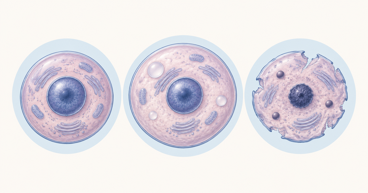
Reversible injury has two classic patterns, acute cell swelling and fatty change. Both appear before the cell reaches the point where recovery is no longer possible.
Cell swelling (hydropic change)
Acute cell swelling is the earliest reversible change and results from the failed Na+/K+ pump described above. The same pathology chapter gives alternative names by tissue: hydropic change in general, ballooning degeneration in the epidermis and cytotoxic oedema in CNS glia.
What you see depends on the level of examination:
- Gross: the organ is heavier, paler and swollen.
- Light microscopy: cells are enlarged and rounded, with pale eosinophilic or finely vacuolated cytoplasm.
- Electron microscopy: swollen mitochondria and endoplasmic reticulum, clumped chromatin, electron-lucent cytosol, ribosomes detached from rough ER, vesiculated ER, membrane whorls (myelin figures), loss of microvilli and surface blebs.
Swelling is reversible up to a point, and it is also the early change in irreversible injury. So swelling alone cannot tell you which way the cell is heading.
Fatty change (steatosis)
Fatty change is the accumulation of lipid in cells, most often described in hepatocytes. It happens when fat import or synthesis exceeds export or degradation. Reduced beta-oxidation or reduced VLDL secretion can both tip the balance.
Grossly, the liver is swollen, yellow and greasy. On microscopy, clear lipid vacuoles push the nucleus to the edge of the cell. The fat itself is dissolved during routine tissue processing, which is why the vacuoles look empty.
Reviews of hepatic steatosis list obesity with insulin resistance, excess alcohol, hepatitis C, drugs and pregnancy-related liver disease among the causes. They also note that triglyceride is not intrinsically toxic, whereas lipotoxic species such as free fatty acids and cholesterol are linked to injury.
Irreversible Cell Injury and the Point of No Return
Irreversible injury is recognised by a cluster of changes rather than a single sign. Swelling increases, lysosomes are disrupted, membranes break down and the nucleus changes severely.
Mitochondrial markers
Failure to restore mitochondrial function marks the shift to irreversible injury in the pathology chapter. On electron microscopy, the mitochondria develop amorphous (flocculent) densities. These densities are considered a marker of irreversibility and probably reflect protein denaturation, as described in studies of ischaemic injury.
Membrane and lysosomal changes
Severe membrane damage lets cell enzymes and proteins leak into the blood, which is why they act as markers of cell death. Lysosomal rupture is also described. However, a tracer study of lysosome stability in lethal injury found that lysosomes released dye only after the cells were already necrotic, so rupture may follow death rather than cause it.
Nuclear changes
The nucleus shows one of three patterns:
- Pyknosis: condensation of chromatin
- Karyorrhexis: fragmentation of the nucleus
- Karyolysis: dissolution of the nucleus
A simple memory aid: the nucleus shrinks (pyknosis), breaks (karyorrhexis) or melts (karyolysis).
Calcification in dead tissue
Dystrophic calcification occurs in dead or dying tissue with a normal serum calcium. Metastatic calcification is the high-calcium form. In dystrophic calcification, mitochondria are the first sites of deposition.
Why there is no single point of no return
Textbook diagrams often draw a clean line between reversible and irreversible injury. The sources do not support a single event. The exact timing and the molecular trigger are not agreed. Practical markers include failure to restore mitochondrial function and ATP, calcium influx, amorphous mitochondrial densities, severe membrane damage and nuclear changes.
Farber's calcium review captures the uncertainty well: either calcium enters after the cell has died, or calcium influx is what makes membrane damage lethal. Both are plausible and neither is proven. Time to irreversibility also varies with metabolic demand, from tens of minutes to hours, so avoid memorising a fixed time unless a question stem gives one.
Reversible vs Irreversible Cell Injury: Comparison Table
The table below pulls the markers from the sections above into one place. To test yourself, cover the right-hand columns and recall them.
| Feature | Reversible injury | Irreversible injury |
|---|---|---|
| Typical patterns | Cell swelling, fatty change | Necrosis |
| Cell size and shape | Enlarged, rounded | Increased swelling, then rupture |
| Cytoplasm (light microscopy) | Pale eosinophilic or finely vacuolated; clear lipid vacuoles in fatty change | Increased eosinophilia in later stages |
| Mitochondria (EM) | Swollen | Amorphous (flocculent) densities |
| Endoplasmic reticulum (EM) | Swollen, vesiculated, ribosomes detached | Further disruption |
| Plasma membrane | Blebs, loss of microvilli, myelin figures | Severe damage, leakage of cell contents |
| Lysosomes | Not described as disrupted | Disruption described |
| Nucleus | Clumped chromatin | Pyknosis, karyorrhexis or karyolysis |
| Outcome | Recovery if the insult is removed | Cell death |
Study note: increased eosinophilia is a general feature of dead cells, so learn it as a supporting clue alongside the nuclear changes rather than as a stand-alone marker.
Types of Necrosis After Cell Injury
Necrosis describes the changes that follow cell death by any mechanism, as Majno and Joris put it. The patterns below are descriptive terms, and StatPearls treats them that way. The pattern depends on the tissue and the cause.
| Type | Key mechanism or feature | Typical setting |
|---|---|---|
| Coagulative | Protein denaturation; tissue architecture persists | Hypoxia and ischaemia; infarcts of kidney, liver, heart, muscle |
| Liquefactive | Dead tissue digested to liquid | Brain and spinal cord; pus-forming infection |
| Caseous | Tissue architecture completely lost | Granulomatous inflammation (granulomas) |
| Fat | Pancreatic lipases; chalky white saponification | Pancreatic injury |
| Gangrenous | Clinical term: dry gangrene is ischaemic coagulative necrosis; wet gangrene is liquefactive after infection | Ischaemic limbs, bowel |
| Fibrinoid | Plasma proteins leak into an injured vessel wall | Vessel walls |
The pathology chapter cited earlier calls the last entry "fibrinoid change", while standard teaching says fibrinoid necrosis. Use the second wording in answers. For a deeper pattern-by-pattern treatment, see this site's post on types of necrosis.
Necrosis vs Apoptosis in Cell Injury
Necrosis and apoptosis differ in cell size, membrane integrity and inflammation. The classic descriptions from the pathology chapter and from Majno and Joris line up as follows.
| Feature | Necrosis | Apoptosis |
|---|---|---|
| Cell size | Swelling | Shrinkage |
| Cell fate | Rupture | Fragmentation into membrane-bound bodies |
| Distribution | Contiguous groups of cells | Single cells |
| Plasma membrane | Disrupted | Intact |
| Nucleus | Karyolysis, pyknosis, karyorrhexis | Fragmented in apoptotic bodies |
| Driver | Accidental in classic descriptions | Caspase-driven |
| Inflammation | Present | Absent |
One nuance is worth holding on to. Regulated forms of necrosis, such as necroptosis, blur the view that necrosis is always accidental, and both processes can coexist in the same tissue.
Ischaemia-Reperfusion Injury: When Restoring Flow Adds Cell Injury
Restoring blood flow to ischaemic tissue can make injury worse. The pathology chapter describes several contributors: oxidative stress, calcium imbalance, opening of the mitochondrial permeability transition pore, endothelial damage, thrombosis and leukocyte infiltration. Damage can continue for several days after reperfusion.
The practical lesson is that "reversible" depends on what happens after the insult as well as during it. A cell that looks salvageable at the moment flow returns may still be lost to the secondary injury.
How to Study Cell Injury for NEET PG
Treat the topic as one causal chain, then attach pictures to each step. A suggested plan:
- Draw the hypoxia chain from memory: ATP loss, pump failure, swelling, calcium entry, enzyme activation.
- Learn reversible markers as a pattern: swelling, blebs, myelin figures, ER changes, fatty change.
- Learn irreversible markers as a pattern: amorphous mitochondrial densities, membrane damage, nuclear changes.
- Rebuild the necrosis and apoptosis tables without looking.
- If a stem gives you a clue such as a brain infarct or a caseating granuloma, think of the matching necrosis type.
As a self-test, explain in two or three sentences, using only the ATP chain, why cell swelling appears before any nuclear change. Then test yourself with questions. The site's cell injury practice questions let you apply the markers to short vignettes.
For mixed practice across subjects, use the NEET PG question bank.
Conclusion
Cell injury is best understood as a sequence: an insult depletes ATP, the pump and membranes fail, and the cell swells. If function returns, the injury is reversible. If mitochondrial function cannot be restored and the membrane and nucleus break down, it is irreversible. Because no single event defines the point of no return, learn the whole set of markers and the reasoning that links them.
Practise this on Reflex
Turn what you just read into recall with 14 years of tagged PYQs.
FAQ
Frequently asked questions
The questions aspirants ask most about this topic.
In reversible injury the cell can recover if the insult is removed. The classic changes are acute cell swelling and fatty change. In irreversible injury mitochondrial function cannot be restored, and the cell shows amorphous mitochondrial densities, severe membrane damage and nuclear changes (pyknosis, karyorrhexis, karyolysis), ending in cell death.
It is the transition from reversible to irreversible injury. No single molecular event or exact time is agreed. Practical markers include failure to restore mitochondrial function and ATP, calcium influx, amorphous mitochondrial densities, severe membrane damage and nuclear changes.
Hypoxia stops oxidative phosphorylation, so ATP falls and the Na+/K+-ATPase fails. Sodium, calcium and water enter the cell while potassium leaks out, and the cell swells. This acute cell swelling is called hydropic change.
Pyknosis is condensation of chromatin, karyorrhexis is fragmentation of the nucleus, and karyolysis is dissolution of the nucleus.
Yes. This is ischaemia-reperfusion injury. Oxidative stress, calcium imbalance, opening of the mitochondrial permeability transition pore, endothelial damage, thrombosis and leukocyte infiltration can all add damage, which may continue for several days.
Stay ahead in your preparation
Get expert tips, exam updates, and high-yield insights delivered straight to your inbox.
No spam. Unsubscribe anytime.
More in Pathology
View all → Pathology
Virchow's Triad for NEET PG: Three Components, Each With Clinical Examples
Virchow's triad lists the three factors that promote thrombosis: endothelial injury, abnormal blood flow and hypercoagulability. Learn each component with clinical examples and a worked renal cell carcinoma case.
 Pathology
Types of Hypersensitivity Reactions: The Gell and Coombs Classification NEET PG Tests Every Cycle
 Pathology
Reed-Sternberg Cells and Hodgkin vs Non-Hodgkin Lymphoma: The Full NEET PG Comparison
 Pathology