Gram Staining for NEET PG: What the Color Actually Tells You About the Cell Wall
By Dr. Utsav Bhattacherjee, MBBS, MBA · 18 Sept 2026 · 12 min read
Last updated: 23 Sept 2026
On this page
Gram staining questions test one thing above everything else: given a described organism, can you predict its Gram stain result from its known cell wall structure, or work the other way and use a stated result to narrow the differential. The stain itself is simple; the value is entirely in connecting it to what it reveals about the organism.
What the Stain Actually Detects
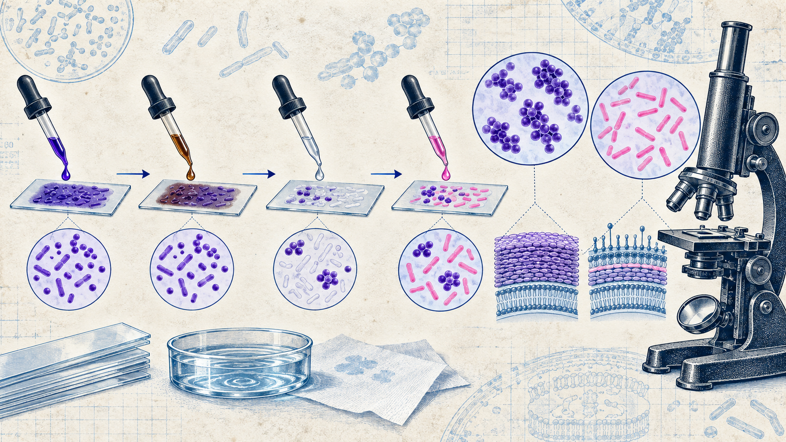
Gram staining differentiates bacteria based on cell wall structure, specifically the thickness of the peptidoglycan layer. Gram-positive organisms have a thick peptidoglycan layer that traps the crystal violet-iodine complex even after decolorization, retaining the purple stain. Gram-negative organisms have a thin peptidoglycan layer sandwiched between an inner membrane and an outer membrane, and the decolorizing step washes the crystal violet out, leaving them to pick up the counterstain, safranin, appearing pink or red instead.
The Four-Step Procedure, and Why Each Step Exists
Crystal violet is applied first, staining all bacteria purple regardless of type, since the dye simply binds broadly at this stage without differentiating anything yet. Iodine is applied second, acting as a mordant that forms a large crystal violet-iodine complex within the cell — this complex is now too large to easily wash out of a thick peptidoglycan layer, but still escapes easily through a thin one.
Decolorization with alcohol or acetone is the actual differentiating step: it dissolves the outer membrane of Gram-negative bacteria, allowing the crystal violet-iodine complex to wash straight out, while the thick, cross-linked peptidoglycan of Gram-positive bacteria holds onto the complex despite the same solvent exposure. This step is also the one most sensitive to technique error — over-decolorizing can make a genuinely Gram-positive organism appear falsely Gram-negative, which is worth knowing as a source of real diagnostic error, not just a procedural detail.
Safranin, the counterstain, is applied last, staining pink or red — but since Gram-positive organisms are already saturated with purple crystal violet, the safranin makes no visible difference to them. Gram-negative organisms, having lost their purple stain during decolorization, pick up this pink counterstain instead, which is what actually makes them visible and distinguishable under the microscope at all.
Named Examples Worth Having Ready
Common Gram-positive organisms worth recognizing on sight include Staphylococcus aureus (clusters), Streptococcus species (chains), and Bacillus and Clostridium species (spore-forming rods). Common Gram-negative organisms include Escherichia coli, Klebsiella, Pseudomonas, and Neisseria species. Building this list mentally against the crystal-violet-versus-safranin logic above turns memorization into recognition — once you know a genus is Gram-negative, you already know it has an outer membrane, thin peptidoglycan, and the clinical implications that follow from that structure.
The Organisms Gram Staining Can't Classify
A meaningful group of clinically important organisms don't fit the Gram-positive or Gram-negative categories at all, and recognizing why is as testable as the staining mechanism itself. Mycobacteria, including the tuberculosis-causing species, have a cell wall rich in mycolic acids that resists the Gram stain's dyes almost entirely — they require an entirely different technique, acid-fast staining, which exploits that same waxy, lipid-rich wall structure rather than working around it.
Mycoplasma species lack a cell wall altogether, having only a cell membrane — since Gram staining depends entirely on peptidoglycan structure to differentiate anything, an organism with no peptidoglycan simply cannot be meaningfully Gram-stained at all. This is also the mechanistic reason antibiotics that target cell wall synthesis, like penicillins, are completely ineffective against Mycoplasma infections, regardless of dose.
Chlamydia species are obligate intracellular organisms with an atypical cell wall that also doesn't take up the Gram stain reliably, and they require cell culture or molecular techniques for identification rather than a simple stained smear. Spirochetes, including the organism causing syphilis, are generally too thin to be reliably visualized by light microscopy with Gram staining at all, requiring dark-field microscopy instead.
Acid-Fast Staining: A Related but Separate Technique
Acid-fast staining itself is worth a brief mechanistic note, since it is frequently confused with Gram staining despite serving a related purpose. Carbol fuchsin is applied with heat to force penetration through the waxy mycolic acid layer, and once inside, this stain resists the acid-alcohol decolorization step that would strip an ordinary Gram stain entirely — hence the name acid-fast. Organisms that retain this stain, most notably Mycobacterium tuberculosis, are described as acid-fast positive, a completely separate classification from Gram-positive or Gram-negative, and one that genuinely requires its own dedicated technique rather than a variant of Gram staining itself.
The Structural Difference Underneath the Color Difference
The staining result is really just a visible proxy for a deeper structural difference worth understanding directly, not just memorizing as a color outcome. Gram-positive cell walls contain teichoic acids embedded within their thick peptidoglycan layer, which are absent in Gram-negative organisms — these teichoic acids play a role in the immune response Gram-positive infections provoke and are part of what the immune system recognizes as a threat signal.
Gram-negative bacteria, in exchange for their thinner peptidoglycan, carry an outer membrane containing lipopolysaccharide (LPS), also called endotoxin. LPS is directly responsible for much of the systemic inflammatory response seen in Gram-negative sepsis — fever, hypotension, and the cascade toward septic shock are driven substantially by the immune system's reaction to LPS release, which is exactly why Gram-negative bloodstream infections carry such a well-established, specific risk of this particular complication compared to many Gram-positive infections.
This outer membrane also explains a separate clinically important property: Gram-negative bacteria are generally more resistant to certain antibiotics and to detergents and dyes, since the outer membrane acts as an additional permeability barrier that Gram-positive organisms simply don't have. A drug that easily penetrates a Gram-positive cell wall may be excluded almost entirely by a Gram-negative outer membrane, which is part of the underlying reason antibiotic selection differs so much between the two categories beyond simply matching a lab result.
Why This Matters Beyond the Stain Itself
Gram stain results directly shape empiric antibiotic selection before culture results are available, which is exactly why this simple, decades-old technique remains central to clinical practice rather than being replaced by more modern methods. A Gram-negative rod seen on a sputum or blood sample points toward organisms like E. coli or Pseudomonas and toward antibiotics effective against that cell wall structure specifically, while a Gram-positive coccus in clusters points toward Staphylococcus and a different empiric choice entirely — the stain result itself is often the very first piece of actionable information a clinician has, hours or days before a full culture and sensitivity result returns.
Reading a Microbiology Vignette Efficiently
A vignette describing a specific stain result, cell wall detail, or antibiotic susceptibility pattern is usually testing whether that detail can be traced back to the Gram-positive or Gram-negative structural distinction first, before narrowing to a specific genus or species. Checking peptidoglycan thickness, presence or absence of an outer membrane, and presence or absence of LPS, in that order, resolves the great majority of these questions before any specific organism needs to be named at all.
A useful summary worth holding as a single mental model: Gram-positive bacteria are structurally simpler on the outside (one thick wall, no outer membrane) but chemically more immunologically active through teichoic acids, while Gram-negative bacteria are structurally more complex (thin wall plus a genuinely separate outer membrane) and carry their major virulence factor, LPS, embedded directly in that additional layer. Every downstream fact about staining behaviour, antibiotic susceptibility, and clinical presentation traces back to this one structural distinction.
For the same mechanism-first classification approach applied to a different topic, see our guide to types of hypersensitivity reactions, and for how much of the paper Microbiology carries overall, our subject-wise weightage breakdown.
Practise this on Reflex
Turn what you just read into recall with 14 years of tagged PYQs.
FAQ
Frequently asked questions
The questions aspirants ask most about this topic.
Alcohol dissolves the outer membrane unique to Gram-negative bacteria, letting the crystal violet-iodine complex wash out through their thin peptidoglycan layer. Gram-positive bacteria's thick, cross-linked peptidoglycan retains the complex despite the same solvent exposure.
Over-decolorizing can strip crystal violet even from Gram-positive organisms, making them falsely appear Gram-negative — a genuine source of diagnostic error, not just a procedural imperfection.
Iodine acts as a mordant, forming a large crystal violet-iodine complex inside the cell. This complex is too large to easily exit a thick peptidoglycan layer, which is exactly the property decolorization then exploits.
Gram-positive bacteria appear purple, retaining the original crystal violet stain. Gram-negative bacteria appear pink or red, having lost the crystal violet during decolorization and picked up the safranin counterstain instead.
LPS, also called endotoxin, is part of the outer membrane and drives much of the systemic inflammatory response in Gram-negative sepsis, including fever, hypotension, and progression toward septic shock.
Mycoplasma lacks a cell wall entirely, having only a cell membrane. Since Gram staining depends on peptidoglycan structure to differentiate organisms, one with no peptidoglycan cannot be meaningfully stained at all.
Acid-fast staining, which exploits the mycolic acid-rich, waxy cell wall of Mycobacteria that resists conventional Gram staining dyes almost entirely.
About the author
Stay ahead in your preparation
Get expert tips, exam updates, and high-yield insights delivered straight to your inbox.
No spam. Unsubscribe anytime.
More in Subject Notes
View all → Microbiology
Spore-Forming Bacteria for NEET PG: Bacillus, Clostridium, and Sterilization
Bacillus vs Clostridium, the spore position clues, and why sterilization protocol depends on this.
 Surgery
Burns for NEET PG: The Rule of Nines, Burn Depth and the Parkland Formula Worked Step by Step
 Radiology
FAST Scan for NEET PG: The Four Windows, eFAST, and What a Positive Result Means
 Dermatology